 PDF
PDF  Views
Views  Share
Share


Isolated bone marrow carcinomatosis: A rare presentation of poorly differentiated adenocarcinoma of the stomach in a young male
CC BY-NC-ND 4.0 · Indian J Med Paediatr Oncol 2016; 37(01): 67
DOI: DOI: 10.4103/0971-5851.177040
Publication History
Article published online:
12 July 2021
© 2016. Indian Society of Medical and Paediatric Oncology. This is an open access article published by Thieme under the terms of the Creative Commons Attribution-NonDerivative-NonCommercial-License, permitting copying and reproduction so long as the original work is given appropriate credit. Contents may not be used for commercial purposes, or adapted, remixed, transformed or built upon. (https://creativecommons.org/licenses/by-nc-nd/4.0/.)
Thieme Medical and Scientific Publishers Pvt. Ltd.
A-12, 2nd Floor, Sector 2, Noida-201301 UP, India
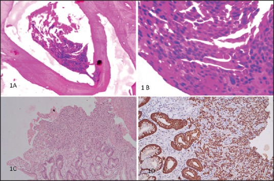
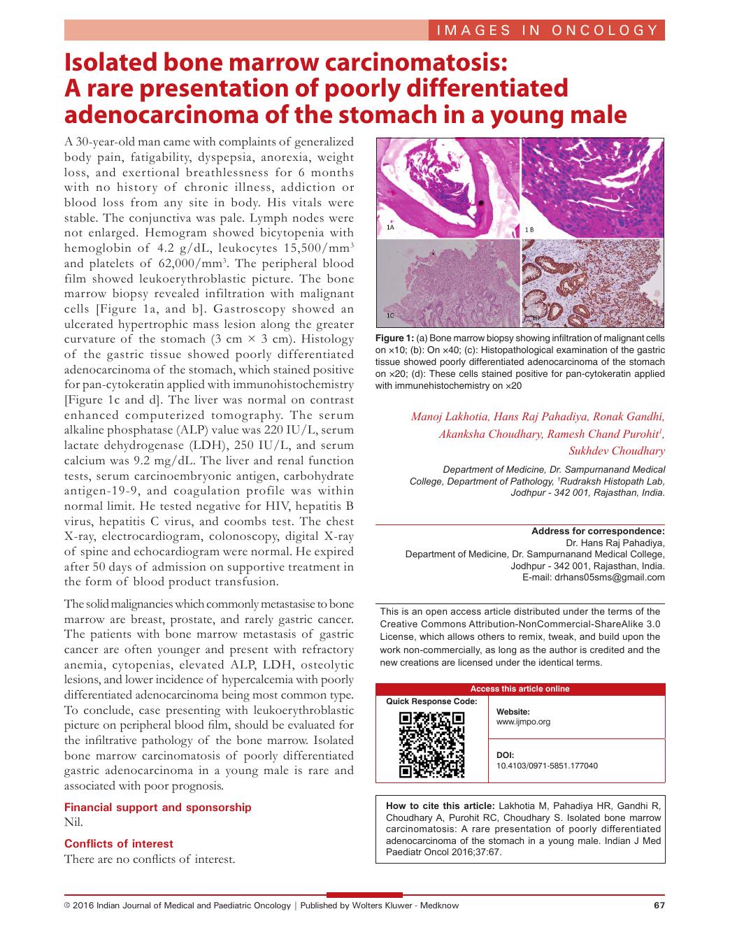
| Fig. 1 (a) Bone marrow biopsy showing infiltration of malignant cells on ×10; (b): On ×40; (c): Histopathological examination of the gastric tissue showed poorly differentiated adenocarcinoma of the stomach on ×20; (d): These cells stained positive for pan-cytokeratin applied with immunehistochemistry on ×20
The solid malignancies which commonly metastasise to bone marrow are breast, prostate, and rarely gastric cancer. The patients with bone marrow metastasis of gastric cancer are often younger and present with refractory anemia, cytopenias, elevated ALP, LDH, osteolytic lesions, and lower incidence of hypercalcemia with poorly differentiated adenocarcinoma being most common type. To conclude, case presenting with leukoerythroblastic picture on peripheral blood film, should be evaluated for the infiltrative pathology of the bone marrow. Isolated bone marrow carcinomatosis of poorly differentiated gastric adenocarcinoma in a young male is rare and associated with poor prognosis.
Financial support and sponsorship
Nil.
Conflicts of interest
There are no conflicts of interest.
| Fig. 1 (a) Bone marrow biopsy showing infiltration of malignant cells on ×10; (b): On ×40; (c): Histopathological examination of the gastric tissue showed poorly differentiated adenocarcinoma of the stomach on ×20; (d): These cells stained positive for pan-cytokeratin applied with immunehistochemistry on ×20