 PDF
PDF  Views
Views  Share
Share


A Patient with Adenocarcinoma Stomach on FLOT (5-Fluorouracil/Leucovorin, Oxaliplatin, and Docetaxel) Chemotherapy Presents with Tooth Discoloration: Spot the Diagnosis
CC BY 4.0 · Indian J Med Paediatr Oncol 2026; 47(01): 070-071
DOI: DOI: 10.1055/s-0045-1813664
Description
A male in his 70s, with no known comorbidities or history of substance abuse, presented with complaints of abdominal discomfort and dyspepsia. Despite being prescribed proton pump inhibitors, his symptoms persisted. An upper gastrointestinal endoscopy revealed a gastric ulcer. Histopathological examination of the biopsy confirmed a diagnosis of moderately differentiated adenocarcinoma stomach. A positron emission tomography PET scan showed hypermetabolic, asymmetrical, circumferential wall nodular thickening involving the medial wall of the antropyloric region of the stomach, along with hypermetabolic enlarged perigastric lymph nodes. The patient was started on perioperative chemotherapy as per the FLOT (5-fluorouracil/leucovorin, oxaliplatin, and docetaxel) protocol. After completion of four cycles of chemotherapy, he underwent surgery. He was then started on postoperative adjuvant chemotherapy.
Following six cycles of chemotherapy, discoloration of the patient's teeth was observed. Specifically, yellow-black discoloration was noted on the lower teeth, and yellowish discoloration on the upper incisors ([Fig. 1]) of the patient. The patient was not taking any additional medications and denied any history of tobacco use. What can be the differential diagnosis?
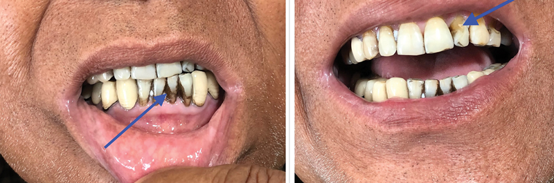
Fig 1: Left: presence of yellow-black discoloration in the lower teeth (arrowhead). Right: yellowish discoloration in the upper incisors (arrowhead).
After completion of a total of eight cycles of chemotherapy as planned, he was then advised to have 3 monthly follow-up. At his 3-month review, the discoloration had partially resolved, implying chemotherapy to be the causative agent. ([Fig. 2]) While chemotherapy-induced tooth discoloration has been previously reported, such occurrences remain relatively rare.[1] [2] [3] Discoloration of teeth and enamel hypoplasia may result from chemotherapeutic agents like vincristine, vinblastine, and cyclophosphamide, which can interfere with ameloblast activity, particularly by disrupting their microtubule-dependent calcium transport systems.[4] This is a rare case of FLOT chemotherapy-induced tooth discoloration. Clinicians must remain vigilant for this adverse effect while administering this chemotherapy regimen to patients.
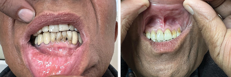
Fig 2 : Left: showing resolution of discoloration in the lower teeth 3 months post last chemotherapy. Right: showing resolution of yellowish discoloration in the upper incisors 3 months post last chemotherapy.
Patient Consent
Patient consent has been taken for this article.
Publication History
Article published online:
01 December 2025
© 2025. The Author(s). This is an open access article published by Thieme under the terms of the Creative Commons Attribution License, permitting unrestricted use, distribution, and reproduction so long as the original work is properly cited. (https://creativecommons.org/licenses/by/4.0/)
Thieme Medical and Scientific Publishers Pvt. Ltd.
A-12, 2nd Floor, Sector 2, Noida-201301 UP, India
We recommend
- A Patient with Adenocarcinoma Stomach on FLOT (5-Fluorouracil/Leucovorin, Oxaliplatin, and Docetaxel) Chemotherapy Presents with Tooth Discoloration: Spot the D...Mayank Kapoor, et al., TH Open, 2025
- A Patient with Adenocarcinoma Stomach on FLOT (5-Fluorouracil/Leucovorin, Oxaliplatin, and Docetaxel) Chemotherapy Presents with Tooth Discoloration: Spot the D...Mayank Kapoor, et al., VCOT Open, 2025
- A Prospective Study to Evaluate the Efficacy of the Fluorouracil, Leucovorin, Oxaliplatin, and Docetaxel Chemotherapy Regimen in Patients with Locally Advanced ...Vijay Kumar Srinivasalu, VCOT Open, 2022
- A Prospective Study to Evaluate the Efficacy of the Fluorouracil, Leucovorin, Oxaliplatin, and Docetaxel Chemotherapy Regimen in Patients with Locally Advanced ...Vijay Kumar Srinivasalu, TH Open, 2022
- Enlightening Diagnosis and Differential Diagnosis of Dental Fluorosis—A Hidden Entity in a CrowdDeepika Patidar, Dental Journal of Advance Studies, 2021
- Targeting MUC1 with fisetin in oral squamous cell carcinomaQian Wang, Genes & Diseases, 2025
- Circulating miR-18b-3p is a novel biomarker predicting chemo-radiotherapy induced oral mucositis in head and neck cancerClaudio Pulito, Signal Transduction and Targeted Therapy, 2025
- First-line camrelizumab (a PD-1 inhibitor) plus apatinib (an VEGFR-2 inhibitor) and chemotherapy for advanced gastric cancer (SPACE): a phase 1 study<svg viewBox="0 0 24 24" fill="none" xmlns="http://www.w3.org/2000/svg">
</svg> Xiaofeng Chen, Signal Transduction and Targeted Therapy, 2024 - Addition of SHR-1701 to first-line capecitabine and oxaliplatin (XELOX) plus bevacizumab for unresectable metastatic colorectal cancerMiao-Zhen Qiu, Signal Transduction and Targeted Therapy, 2024
- PD-1 antibody camrelizumab plus apatinib and SOX as first-line treatment in patients with AFP-producing gastric or gastro-esophageal junction adenocarcinoma (CA...Yakun WANG, Signal Transduction and Targeted Therapy, 2025
Description
A male in his 70s, with no known comorbidities or history of substance abuse, presented with complaints of abdominal discomfort and dyspepsia. Despite being prescribed proton pump inhibitors, his symptoms persisted. An upper gastrointestinal endoscopy revealed a gastric ulcer. Histopathological examination of the biopsy confirmed a diagnosis of moderately differentiated adenocarcinoma stomach. A positron emission tomography PET scan showed hypermetabolic, asymmetrical, circumferential wall nodular thickening involving the medial wall of the antropyloric region of the stomach, along with hypermetabolic enlarged perigastric lymph nodes. The patient was started on perioperative chemotherapy as per the FLOT (5-fluorouracil/leucovorin, oxaliplatin, and docetaxel) protocol. After completion of four cycles of chemotherapy, he underwent surgery. He was then started on postoperative adjuvant chemotherapy.
Following six cycles of chemotherapy, discoloration of the patient's teeth was observed. Specifically, yellow-black discoloration was noted on the lower teeth, and yellowish discoloration on the upper incisors ([Fig. 1]) of the patient. The patient was not taking any additional medications and denied any history of tobacco use. What can be the differential diagnosis?
Fig 1: Left: presence of yellow-black discoloration in the lower teeth (arrowhead). Right: yellowish discoloration in the upper incisors (arrowhead).
After completion of a total of eight cycles of chemotherapy as planned, he was then advised to have 3 monthly follow-up. At his 3-month review, the discoloration had partially resolved, implying chemotherapy to be the causative agent. ([Fig. 2]) While chemotherapy-induced tooth discoloration has been previously reported, such occurrences remain relatively rare.[1] [2] [3] Discoloration of teeth and enamel hypoplasia may result from chemotherapeutic agents like vincristine, vinblastine, and cyclophosphamide, which can interfere with ameloblast activity, particularly by disrupting their microtubule-dependent calcium transport systems.[4] This is a rare case of FLOT chemotherapy-induced tooth discoloration. Clinicians must remain vigilant for this adverse effect while administering this chemotherapy regimen to patients.
Fig 2 : Left: showing resolution of discoloration in the lower teeth 3 months post last chemotherapy. Right: showing resolution of yellowish discoloration in the upper incisors 3 months post last chemotherapy.
Acknowledgment
The manuscript has been read and approved by all the authors, and the requirements for authorship as stated earlier in this document have been met, and each author believes that the manuscript represents honest work.
Patient Consent
Patient consent has been taken for this article.
- Busenhart DM, Erb J, Rigakos G, Eliades T, Papageorgiou SN. Adverse effects of chemotherapy on the teeth and surrounding tissues of children with cancer: a systematic review with meta-analysis. Oral Oncol 2018; 83: 64-72
- Poulopoulos A, Papadopoulos P, Andreadis D. Chemotherapy: oral side effects and dental interventions -a review of the literature. Stomatol Dis Sci 2017; 1: 35-49
- Oral Complications of Chemotherapy and Head/Neck Radiation. (PDQ)–Patient Version - NCI. May 23, 2022. Accessed February 24, 2023 at: https://www.cancer.gov/about-cancer/treatment/side-effects/mouth-throat/oral-complications-pdq
- Oğuz A, Cetiner S, Karadeniz C, Alpaslan G, Alpaslan C, Pinarli G. Long-term effects of chemotherapy on orodental structures in children with non-Hodgkin's lymphoma. Eur J Oral Sci 2004; 112 (01) 8-11
References
Address for correspondence
Publication History
Article published online:
01 December 2025
© 2025. The Author(s). This is an open access article published by Thieme under the terms of the Creative Commons Attribution License, permitting unrestricted use, distribution, and reproduction so long as the original work is properly cited. (https://creativecommons.org/licenses/by/4.0/)
Thieme Medical and Scientific Publishers Pvt. Ltd.
A-12, 2nd Floor, Sector 2, Noida-201301 UP, India
We recommend
- A Patient with Adenocarcinoma Stomach on FLOT (5-Fluorouracil/Leucovorin, Oxaliplatin, and Docetaxel) Chemotherapy Presents with Tooth Discoloration: Spot the D...Mayank Kapoor, et al., TH Open, 2025
- A Patient with Adenocarcinoma Stomach on FLOT (5-Fluorouracil/Leucovorin, Oxaliplatin, and Docetaxel) Chemotherapy Presents with Tooth Discoloration: Spot the D...Mayank Kapoor, et al., VCOT Open, 2025
- A Prospective Study to Evaluate the Efficacy of the Fluorouracil, Leucovorin, Oxaliplatin, and Docetaxel Chemotherapy Regimen in Patients with Locally Advanced ...Vijay Kumar Srinivasalu, VCOT Open, 2022
- A Prospective Study to Evaluate the Efficacy of the Fluorouracil, Leucovorin, Oxaliplatin, and Docetaxel Chemotherapy Regimen in Patients with Locally Advanced ...Vijay Kumar Srinivasalu, TH Open, 2022
- Enlightening Diagnosis and Differential Diagnosis of Dental Fluorosis—A Hidden Entity in a CrowdDeepika Patidar, Dental Journal of Advance Studies, 2021
- Photodynamic therapy in the treatment of the right buccal mucosa verrucous carcinoma: a case report and literature reviewLV Shiping, Journal of Prevention and Treatment for Stomatological Diseases , 2024
- Chemotherapy for Esophageal, Gastric and Colorectal Cancers<svg viewBox="0 0 24 24" fill="none" xmlns="http://www.w3.org/2000/svg">
</svg> The Medical Letter, 2006 - Comparison of Clinical Outcomes of Borderline Resectable Pancreatic Cancer According to the Neoadjuvant Chemo-Regimens: Gemcitabine versus FOLFIRINOXYoo Jin Choi, Gut And Liver
- Lipomatosis: An Unusual Side-effect of Cytotoxic Chemotherapy?<svg viewBox="0 0 24 24" fill="none" xmlns="http://www.w3.org/2000/svg">
</svg> Cronin, Patricia A., Acta Dermato-Venereologica, 2010 - In Brief: Trifluridine/Tipiracil (Lonsurf) for Metastatic Colorectal Cancer (online only)<svg viewBox="0 0 24 24" fill="none" xmlns="http://www.w3.org/2000/svg">
</svg> The Medical Letter, 2016
Fig 1: Left: presence of yellow-black discoloration in the lower teeth (arrowhead). Right: yellowish discoloration in the upper incisors (arrowhead).
Fig 2 : Left: showing resolution of discoloration in the lower teeth 3 months post last chemotherapy. Right: showing resolution of yellowish discoloration in the upper incisors 3 months post last chemotherapy.
- Busenhart DM, Erb J, Rigakos G, Eliades T, Papageorgiou SN. Adverse effects of chemotherapy on the teeth and surrounding tissues of children with cancer: a systematic review with meta-analysis. Oral Oncol 2018; 83: 64-72
- Poulopoulos A, Papadopoulos P, Andreadis D. Chemotherapy: oral side effects and dental interventions -a review of the literature. Stomatol Dis Sci 2017; 1: 35-49
- Oral Complications of Chemotherapy and Head/Neck Radiation. (PDQ)–Patient Version - NCI. May 23, 2022. Accessed February 24, 2023 at: https://www.cancer.gov/about-cancer/treatment/side-effects/mouth-throat/oral-complications-pdq
- Oğuz A, Cetiner S, Karadeniz C, Alpaslan G, Alpaslan C, Pinarli G. Long-term effects of chemotherapy on orodental structures in children with non-Hodgkin's lymphoma. Eur J Oral Sci 2004; 112 (01) 8-11