 PDF
PDF  Views
Views  Share
Share


A Rare Case of Hemangioendothelioma of Urinary Bladder
CC BY-NC-ND 4.0 · Indian J Med Paediatr Oncol 2017; 38(01): 65-66
DOI: DOI: 10.4103/ijmpo.ijmpo_123_16
Abstract
Hemangioendothelioma is a vascular tumor of endothelial nature of intermediate grade. It most commonly arises from soft tissue of upper and lower extremities. We report a rare case of epithelioid hemangioendothelioma of the urinary bladder. Histologically, it was a vascular tumor formed by smaller capillaries lined by plump epithelioid cells having eosinophilic cytoplasm. Diagnosis was confirmed by immunohistochemistry, as the tumor cells were positive for CD34 and smooth muscle actin.
Publication History
Article published online:
06 July 2021
© 2017. Indian Society of Medical and Paediatric Oncology. This is an open access article published by Thieme under the terms of the Creative Commons Attribution-NonDerivative-NonCommercial-License, permitting copying and reproduction so long as the original work is given appropriate credit. Contents may not be used for commercial purposes, or adapted, remixed, transformed or built upon. (https://creativecommons.org/licenses/by-nc-nd/4.0/.)
Thieme Medical and Scientific Publishers Pvt. Ltd.
A-12, 2nd Floor, Sector 2, Noida-201301 UP, India
Abstract
Hemangioendothelioma is a vascular tumor of endothelial nature of intermediate grade. It most commonly arises from soft tissue of upper and lower extremities. We report a rare case of epithelioid hemangioendothelioma of the urinary bladder. Histologically, it was a vascular tumor formed by smaller capillaries lined by plump epithelioid cells having eosinophilic cytoplasm. Diagnosis was confirmed by immunohistochemistry, as the tumor cells were positive for CD34 and smooth muscle actin.
Introduction
Hemangioendothelioma is a vascular tumor of endothelial nature of intermediate grade. Tumors included in this group have the ability to recur locally and have some ability to metastasize but at a far reduced level compared to angiosarcoma.[1] Vascular neoplasms are uncommon in urinary bladder, especially there are only two reported cases of epithelioid hemangioendothelioma (EHE) of urinary bladder after extensive search.[2,3] We describe a rare case of EHE of the urinary bladder, in which was confirmed by histopathology followed by immunohistochemistry (IHC).
Case Report
A 48-year-old male presented with a history of episodic hematuria for the past 8 years. He was nondiabetic, normotensive, and euthyroid. No definite diagnosis and treatment was done for the past 8 years. On clinical examination, no abnormality was detected. Routine hematological and biochemical parameters were within the normal limit. This patient's renal parameters (blood urea and serum creatinine) were within normal limits at presentation. He had no history of smoking, medicinal exposure, occupational exposure, or any chronic infection of bladder, which are considered as significant risk factors for bladder tumor. His urine cytology on multiple occasions revealed markedly high red blood cell (RBC) count and occasional blood clot. There was no evidence of any malignant cells.
He was evaluated with ultrasound (USG) of kidney, ureter, and bladder (KUB) region and computed tomography (CT) scan of the abdomen. In USG, presence of a mass lesion was suggested at the left side of pelvis in the line of ureter. CT scan of KUB revealed a 2.2 cm × 2 cm enhancing calcified nodule attached to urinary bladder dome [Figure 1].
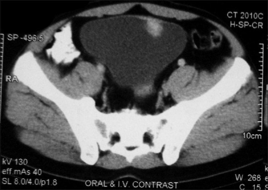
| Figure 1Contrast-enhanced computed tomography of the bladder showing an enhancing mass lesion involving the dome of the urinary bladder
The patient was posted for cystoscopy, and during operation, solitary >2 cm solid raspberry-like highly vascular tumor was found at the dome of the bladder. A transurethral complete resection of the tumor was done by cystoscopy with biopsies from deeper tissue. Rest of the bladder was devoid of any abnormality.
Histopathological examination was carried out on formalin-fixed, paraffin-embedded tissue sections prepared from biopsy specimen. On histopathological examination, sections studied showed a vascular tumor formed by smaller capillaries lined by plump epithelioid cells having eosinophilic cytoplasm. Some of the individual cells showed a vascular lumen containing RBCs. Lining transitional epithelium was unremarkable. No evidence of malignancy was detected [Figure 2]. Immunostaining was performed by applying the streptavidin–biotin–peroxidase conjugate method in an automated stainer (Ventana BenchMark XT, Ventana Medical Systems, Rosche) utilizing the manufacturer's protocol with prediluted ready-to-use antibodies (Dako, Glostrup, Denmark). On IHC, tumor cells were positive for CD34 and smooth muscle actin but negative for CK, EMA, CD-68, S-100, vimentin, and CD56 [Figure 2]. These features are consistent with EHE, ruling out possibility of other soft tissue and epithelial neoplasms. At 6-month follow-up, the patient was asymptomatic, and on USG, there was no recurrence in the bladder.
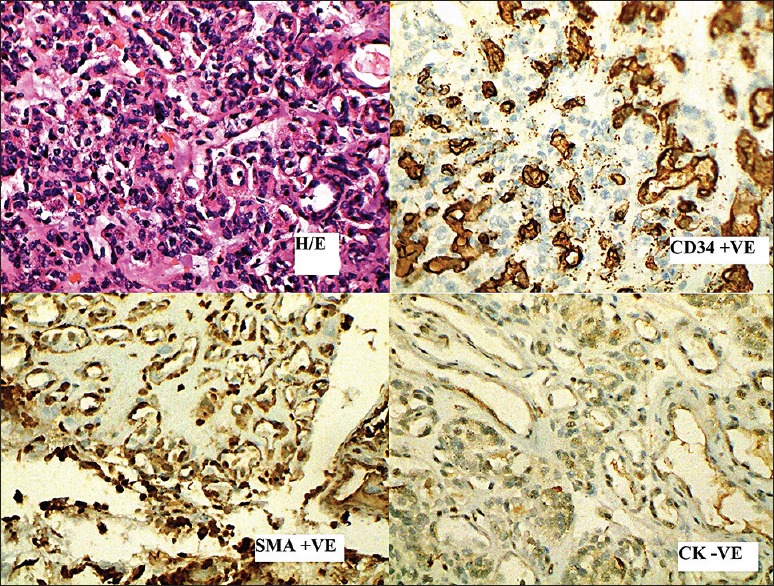
| Figure 2Photograph shows routine section of proliferation of plump endothelial cells (H and E, ×400) and positivity for immunostain CD34, smooth muscle actin, and negative immunostain CK (×400)
Discussion
EHE is a vascular tumor of adults which is characterized by an “epithelioid” or “histiocytoid” endothelial cell. They involve both superficial and deep soft tissue. They are characterized by rounded to slightly spindled endothelial cells, having eosinophilic cytoplasm with vacuolization with rounded nuclei. The behavior of EHE is intermediate between hemangiomas and conventional (high grade) angiosarcomas.[4,5,6] While EHE involves commonly the soft tissue of the extremities, visceral involvement may also occur.[7] The differential diagnoses that were considered based on the histopathological characteristic are granular cell tumor, inflammatory myofibroblastic tumor, EHE, and epithelioid leiomyosarcoma.
References
- Deyrup AT, Tighiouart M, Montag AG, Weiss SW. Epithelioid hemangioendothelioma of soft tissue: A proposal for risk stratification based on 49 cases. Am J Surg Pathol 2008;32:924-7.
- Gupta NP, Kolla SB, Panda S, Sharma MC. Epitheloid hemangioendothelioma of urinary bladder. Indian J Urol 2008;24:253-5.
- Geramizadeh B, Banani A, Foroutan H, Aminsharifi A, Karimi M. Malignant epithelioid hemangioendothelioma of the bladder: The first case report in a child. J Pediatr Surg 2009;44:1443-5.
- Weiss SW, Enzinger FM. Epithelioid hemangioendothelioma: A vascular tumor often mistaken for a carcinoma. Cancer 1982;50:970-81.
- Weiss SW, Ishak KG, Dail DH, Sweet DE, Enzinger FM. Epithelioid hemangioendothelioma and related lesions. Semin Diagn Pathol 1986;3:259-87.
- Mentzel T, Beham A, Calonje E, Katenkamp D, Fletcher CD. Epithelioid hemangioendothelioma of skin and soft tissues: Clinicopathologic and immunohistochemical study of 30 cases. Am J Surg Pathol 1997;21:363-74.
- Läuffer JM, Zimmermann A, Krähenbühl L, Triller J, Baer HU. Epithelioid hemangioendothelioma of the liver. A rare hepatic tumor. Cancer 1996;78:2318-27.
| Figure 1Contrast-enhanced computed tomography of the bladder showing an enhancing mass lesion involving the dome of the urinary bladder
| Figure 2Photograph shows routine section of proliferation of plump endothelial cells (H and E, ×400) and positivity for immunostain CD34, smooth muscle actin, and negative immunostain CK (×400)
References
- Deyrup AT, Tighiouart M, Montag AG, Weiss SW. Epithelioid hemangioendothelioma of soft tissue: A proposal for risk stratification based on 49 cases. Am J Surg Pathol 2008;32:924-7.
- Gupta NP, Kolla SB, Panda S, Sharma MC. Epitheloid hemangioendothelioma of urinary bladder. Indian J Urol 2008;24:253-5.
- Geramizadeh B, Banani A, Foroutan H, Aminsharifi A, Karimi M. Malignant epithelioid hemangioendothelioma of the bladder: The first case report in a child. J Pediatr Surg 2009;44:1443-5.
- Weiss SW, Enzinger FM. Epithelioid hemangioendothelioma: A vascular tumor often mistaken for a carcinoma. Cancer 1982;50:970-81.
- Weiss SW, Ishak KG, Dail DH, Sweet DE, Enzinger FM. Epithelioid hemangioendothelioma and related lesions. Semin Diagn Pathol 1986;3:259-87.
- Mentzel T, Beham A, Calonje E, Katenkamp D, Fletcher CD. Epithelioid hemangioendothelioma of skin and soft tissues: Clinicopathologic and immunohistochemical study of 30 cases. Am J Surg Pathol 1997;21:363-74.
- Läuffer JM, Zimmermann A, Krähenbühl L, Triller J, Baer HU. Epithelioid hemangioendothelioma of the liver. A rare hepatic tumor. Cancer 1996;78:2318-27.