 PDF
PDF  Views
Views  Share
Share


Ewing’s Sarcoma of the Calcaneum
CC BY-NC-ND 4.0 · Indian J Med Paediatr Oncol 2017; 38(04): 542-544
DOI: DOI: 10.4103/ijmpo.ijmpo_50_16
Abstract
Ewing's sarcoma of the calcaneum is rare. Radiological features of this tumor can be misinterpreted as other benign bone tumors due to its rarity. The overall prognosis of Ewing's sarcoma of calcaneum is inferiorcompared to other sites of this tumor. Hence, these tumors should have extensive radiological evaluation and histological confirmation as misdiagnosis and treatment delays will have detrimental outcomes.
Publication History
Article published online:
04 July 2021
© 2017. Indian Society of Medical and Paediatric Oncology. This is an open access article published by Thieme under the terms of the Creative Commons Attribution-NonDerivative-NonCommercial-License, permitting copying and reproduction so long as the original work is given appropriate credit. Contents may not be used forcommercial purposes, or adapted, remixed, transformed or built upon. (https://creativecommons.org/licenses/by-nc-nd/4.0/.)
Thieme Medical and Scientific Publishers Pvt. Ltd.
A-12, 2nd Floor, Sector 2, Noida-201301 UP, India
Abstract
Ewing's sarcoma of the calcaneum is rare. Radiological features of this tumor can be misinterpreted as other benign bone tumors due to its rarity. The overall prognosis of Ewing's sarcoma of calcaneum is inferior compared to other sites of this tumor. Hence, these tumors should have extensive radiological evaluation and histological confirmation as misdiagnosis and treatment delays will have detrimental outcomes.
Ewing sarcoma is the second most common bone tumor occurring in children. It usually affects children of 5–20 years of age. This tumor occurring in the small bones of the hand and feet is rare. We report a case of Ewing's sarcoma of the calcaneum in a 10-year-old girl.
A 10-year-old girl presented to an orthopedic center with pain and swelling of the left heel of 2 months duration. Physical examination revealed a firm immobile tender swelling in the left heel. Distal neurovascular status was normal. No significant medical history was noted.
Radiograph of the left heel showed an expansile lytic lesion in the calcaneum with cortical break and no periosteal reaction [Figure 1]. Magnetic resonance imaging (MRI) of the ankle revealed well-defined expansile cystic lesion in the calcaneum. The lesion appears predominantly isointense on T1-weighted imaging and heterogeneously hyperintense in T2-weighted imaging with septations and fluid layering [Figure 2]. There is a breach in the medial aspect with the extension of the lesion into the adjacent soft tissue with enhancement on postcontrast study. The radiologic impression was of aneurysmal bone cyst.
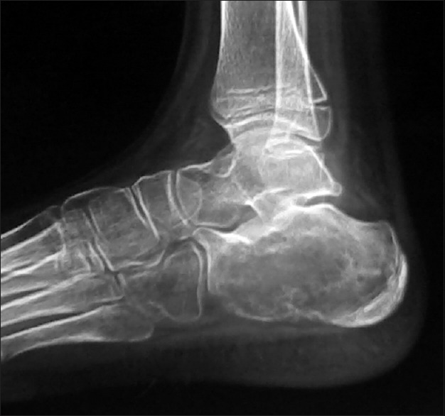
| Figure 1:Radiograph showing well-defined expansile lytic lesion in the calcaneum
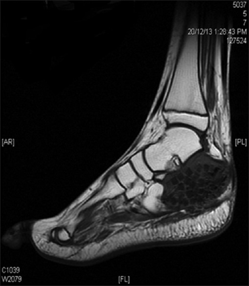
| Figure 2:On T1-weighted imaging, the lesion was seen as low intensity with bone grafts
Curettage with bone grafting was performed at the orthopedic center. Histology of the lesion revealed highly cellular tumor arranged in lobules with foci of cartilage and underlying bone trabeculae infiltrated by compact sheets of round cells with negligible cytoplasm and hyperchromatic nuclei with indistinct nucleoli. Osteoid production was not seen.
She was referred for further management to our center. A review of the slides confirmed a malignant round-cell tumor suggestive of Ewing's sarcoma. Immunohistochemistry showed diffuse strong membrane positivity to CD99 and negativity to the left coronary artery. Computed tomography (CT) of the chest, bone scan, and bone marrow examination excluded the presence of distant metastases. Diagnosis of nonmetastatic Ewing's sarcoma of the calcaneum was made.
After preoperative chemotherapy with alternating cycles of VDC (vincristine/doxorubicin/cyclophosphamide) and ifosfamide/etoposide (IE) for 12 weeks, she underwent below-knee amputation. Then she completed adjuvant chemotherapy with remaining cycles of VDC/IE chemotherapy and is presently on follow-up.
Ewing's sarcoma is the second most common bone tumor occurring in children between the ages of 5 and20 years. It affects the diaphysis of the long bones and is less commonly seen in flat bones and rarely in small bones of the arm and feet.[1] The only prognostic features were tumour site and treatment according to study by casadei et al.[2] Cook has reported 29 cases of Ewing's sarcoma of the calcaneum.[3] They present as swelling of the bone or soft tissue accompanied with pain and sometimes fever.
The radiographic features of Ewing's sarcoma include lytic, condensed lesion with permeative margins, cortical destruction, and aggressive periosteal reaction. There is generally an extraosseous soft-tissue component. However, in our patient, there was an expansile lytic lesion with no periosteal reaction or soft-tissue component, and an initial diagnosis of the aneurysmal bone cyst was made. Cystic changes with absence of cortical thickening and lamellated or speculated periosteal reaction are commonly seen in Ewing's sarcoma of the hand and feet.[4] MRI can help in differentiating the tumor, but definitive diagnosis can be established only with biopsy. Skip metastases are commonly seen in the adjacent bones.[5,6]
Even with clinical and radiological findings, Ewing's sarcoma can be misinterpreted as osteomyelitis, cartilaginous tumor, aneurysmal bone cysts, giant cell lesion, lymphoma, or osteosarcoma, and the distinction often requires extensive evaluation using varied imaging modalities.
The classical histology of Ewing's sarcoma is composed of sheets of small cells with high nuclear to cytoplasmic ratio. There are scant eosinophilic cytoplasm and round nuclei with finely dispersed chromatin and one or more tiny nucleoli. Immunohistochemistry plays an integral part in distinguishing these tumors from the other round cell tumors.[7] The classic Ewing's sarcoma cells show membranous expression of CD99 and nuclear staining of FLI1, and as per the degree of neuroectodermal differentiation, there may be an expression of neuron specific enolase, synaptophysin, and S100 protein.[8] Histologically, two patterns of presentation of Ewing's sarcoma of calcaneum –the diffuse and filigree type – have been described, with the diffuse pattern having better prognosis than the filigree pattern.[10] The Ewing's family of tumors is associated with translocation t (11; 22) (q24; q12) in 85% of cases. Due to financial constraints, FL1 and further genetic testing could not be done in our case.
Neoadjuvant chemotherapy followed by surgery is the mainstay of treatment. Radiation as a local form of treatment had worst outcomes in terms of morbidities and survival; hence, surgery is the best form of local therapy.[9] Radiation is indicated if there is a residual tumor after surgical excision. Tumor necrosis after neoadjuvant chemotherapy is an independent prognostic marker.[10] The survival rates of Ewing's sarcoma have improved with multimodality treatment. Prognostic factors influencing outcomes in Ewing's sarcoma are age, size, site, stage, and response to treatment. Extremity tumors have better prognosis compared to pelvic tumor and extraskeletal tumors. Ewing's sarcomas of the small bones of the hand and feet are rare and have poor outcomes, and among them, the calcaneal tumors have the worst prognosis.
Ewing's sarcoma of calcaneum is rare and has poor outcomes compared to tumors of other small bones. This case report reaffirms that the routine radiological management of Ewing's sarcoma should include radiography and MRI of the affected region, along with bone scan and CT of the chest. Histological confirmation is the mainstay of diagnosis and differentiates it from other benign pathologies. A multimodality approach is the mainstay of treatment. It is necessary to be aware of unusual locations of Ewing sarcoma for early diagnosis and better outcomes.
Financial support and sponsorship
Nil.
Conflicts of interest
There are no conflicts of interest.
References
- Adkins CD, Kitaoka HB, Seidl RK, Pritchard DJ. Ewing's sarcoma of the foot. Clin Orthop Relat Res 1997;(343):173-82.
- Casadei R, Magnani M, Biagini R, Mercuri M. Prognostic factors in Ewing's sarcoma of the foot. Clin Orthop Relat Res 2004;(420):230-8.
- Cook MA, Manfredi OL. Ewing's sarcoma of the hand: A case report. Bull Hosp Jt Dis 1996;55:75-7.
- Reinus WR, Gilula LA, Shirley SK, Askin FB, Siegal GP. Radiographic appearance of Ewing sarcoma of the hands and feet: Report from the intergroup Ewing sarcoma study. AJR Am J Roentgenol 1985;144:331-6.
- Dahlin DC, Coventry MB, Scanlon PW. Ewing's sarcoma. A critical analysis of 165 cases. J Bone Joint Surg Am 1961;43-A:185-92.
- Jalal H, Belhadj Z, Enneddam H, Madhar M, Fikry T, Essadki O, et al. Contribution of magnetic resonance imaging in the diagnosis of talus skip metastases of Ewing's sarcoma of the calcaneus in a child: A case report. J Med Case Rep 2011;5:451.
- Shirley SK, Askin FB, Gilula LA, Vietti TJ, Thomas PR, Siegal GP, et al. Ewing's sarcoma in bones of the hands and feet: A clinicopathologic study and review of the literature. J Clin Oncol 1985;3:686-97.
- Desai SS, Jambhekar NA. Pathology of Ewing's sarcoma/PNET: Current opinion and emerging concepts. Indian J Orthop 2010;44:363-8.
- Escobedo EM, Bjorkengren AG, Moore SG. Ewing's sarcoma of the hand. AJR Am J Roentgenol 1992;159:101-2.
- ;Picci P, Böhling T, Bacci G, Ferrari S, Sangiorgi L, Mercuri M, et al. Chemotherapy induced tumor necrosis as a prognostic factor in localized Ewing's sarcoma of the extremities. J Clin Oncol 1997;15:1553-9.
| Figure 1:Radiograph showing well-defined expansile lytic lesion in the calcaneum
| Figure 2:On T1-weighted imaging, the lesion was seen as low intensity with bone grafts
References
- Adkins CD, Kitaoka HB, Seidl RK, Pritchard DJ. Ewing's sarcoma of the foot. Clin Orthop Relat Res 1997;(343):173-82.
- Casadei R, Magnani M, Biagini R, Mercuri M. Prognostic factors in Ewing's sarcoma of the foot. Clin Orthop Relat Res 2004;(420):230-8.
- Cook MA, Manfredi OL. Ewing's sarcoma of the hand: A case report. Bull Hosp Jt Dis 1996;55:75-7.
- Reinus WR, Gilula LA, Shirley SK, Askin FB, Siegal GP. Radiographic appearance of Ewing sarcoma of the hands and feet: Report from the intergroup Ewing sarcoma study. AJR Am J Roentgenol 1985;144:331-6.
- Dahlin DC, Coventry MB, Scanlon PW. Ewing's sarcoma. A critical analysis of 165 cases. J Bone Joint Surg Am 1961;43-A:185-92.
- Jalal H, Belhadj Z, Enneddam H, Madhar M, Fikry T, Essadki O, et al. Contribution of magnetic resonance imaging in the diagnosis of talus skip metastases of Ewing's sarcoma of the calcaneus in a child: A case report. J Med Case Rep 2011;5:451.
- Shirley SK, Askin FB, Gilula LA, Vietti TJ, Thomas PR, Siegal GP, et al. Ewing's sarcoma in bones of the hands and feet: A clinicopathologic study and review of the literature. J Clin Oncol 1985;3:686-97.
- Desai SS, Jambhekar NA. Pathology of Ewing's sarcoma/PNET: Current opinion and emerging concepts. Indian J Orthop 2010;44:363-8.
- Escobedo EM, Bjorkengren AG, Moore SG. Ewing's sarcoma of the hand. AJR Am J Roentgenol 1992;159:101-2.
- ;Picci P, Böhling T, Bacci G, Ferrari S, Sangiorgi L, Mercuri M, et al. Chemotherapy induced tumor necrosis as a prognostic factor in localized Ewing's sarcoma of the extremities. J Clin Oncol 1997;15:1553-9.