 PDF
PDF  Views
Views  Share
Share


Fatal hematogenous relapse of mucinous borderline ovarian tumor of intestinal type
CC BY-NC-ND 4.0 · Indian J Med Paediatr Oncol 2013; 34(02): 134-135
DOI: DOI: 10.4103/0971-5851.116222
Abstract
We describe an unusual case of fatal hematogenous relapse of borderline mucinous ovarian tumour of intestinal type after three years of primary optimal cytoreduction with dissemination to liver, bones and lymphangitic pattern of spread in lungs with resistance to standard chemotherapy.
Publication History
Article published online:
20 July 2021
© 2013. Indian Society of Medical and Paediatric Oncology. This is an open access article published by Thieme under the terms of the Creative Commons Attribution-NonDerivative-NonCommercial-License, permitting copying and reproduction so long as the original work is given appropriate credit. Contents may not be used for commercial purposes, or adapted, remixed, transformed or built upon. (https://creativecommons.org/licenses/by-nc-nd/4.0/.)
Thieme Medical and Scientific Publishers Pvt. Ltd.
A-12, 2nd Floor, Sector 2, Noida-201301 UP, India
Abstract
We describe an unusual case of fatal hematogenous relapse of borderline mucinous ovarian tumour of intestinal type after three years of primary optimal cytoreduction with dissemination to liver, bones and lymphangitic pattern of spread in lungs with resistance to standard chemotherapy.
A 61-year-old lady who has beendiagnosed as pathological stage IA (limited to one ovary with no capsule breach) borderline ovarian tumor of intestinal type [Figure 1] in 2008 has come to us three years later with abdominal pain, backpain and exertional breathlessness. Computed tomographyevaluation showed liverlesion, extensive lymphangitic pulmonary lesion and scattered vertebral lesion but noperitoneal disease [Figure 2]. Bonescan showed multiple uptakes in axial and appendicular skeleton. Biopsy from liver lesion showed mucin positive adenocarcinoma consistent with ovarian origin positive for cytokeratin 7, cytokeratin 20 CK7, CK20) and cancer antigen 125 (CA 125) and negative for caudal type homeobox 2 (CDX2) [Figures [Figures33 and and4].4]. Her upper and lower Gastrointestinal endoscopies were normal. She had progressive disease despite paclitaxel and carboplatin based therapy and failed subsequent secondline therapy before she eventually died of disease related respiratory failure.
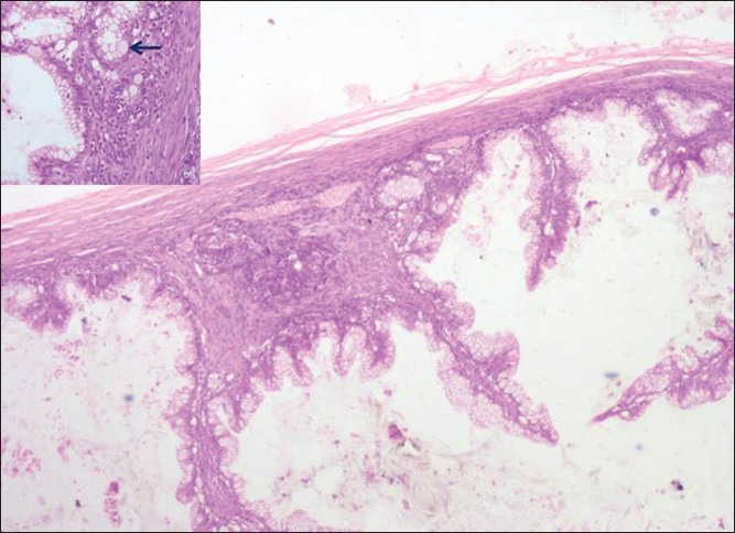
| Fig. 1 Section showing borderline mucinous tumor-intestinal type withgoblet cells (black arrow) of ovary (H and E, ×40) and inset (H and E, ×200)
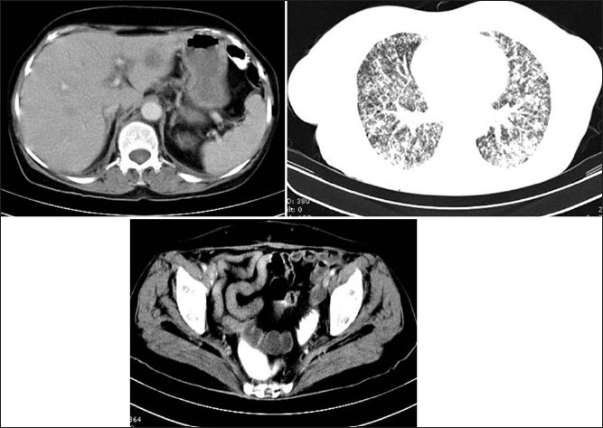
| Fig. 2 Computed tomography evaluation showed liver lesion and extensive lymphangitic pulmonary lesion and scattered vertebral lesion but no peritoneal disease
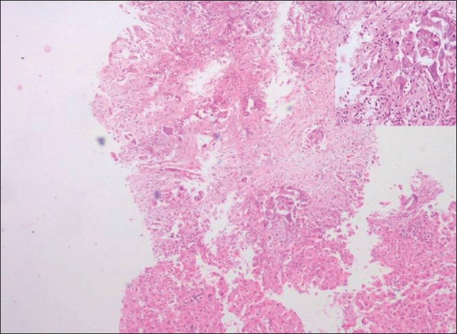
| Fig. 3 Section showing metastatic deposits of mucinous adenocarcinoma (H and E, ×40) and inset (H and E, ×200)
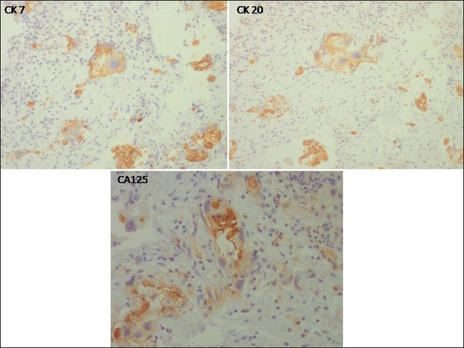
| Fig. 4 Special immunostains highlight immunopisitivity for Ck 7, Ck 20 and Ca125. (Dab, ×200)
Footnotes
Source of Support: Nil
Conflict of Interest: None declared.
| Fig. 1 Section showing borderline mucinous tumor-intestinal type withgoblet cells (black arrow) of ovary (H and E, ×40) and inset (H and E, ×200)
| Fig. 2 Computed tomography evaluation showed liver lesion and extensive lymphangitic pulmonary lesion and scattered vertebral lesion but no peritoneal disease
| Fig. 3 Section showing metastatic deposits of mucinous adenocarcinoma (H and E, ×40) and inset (H and E, ×200)
| Fig. 4 Special immunostains highlight immunopisitivity for Ck 7, Ck 20 and Ca125. (Dab, ×200)