 PDF
PDF  Views
Views  Share
Share


Fibroepithelial Polyp with Sebaceous Hyperplasia: A Case Report
CC BY-NC-ND 4.0 · Indian J Med Paediatr Oncol 2017; 38(03): 404-406
DOI: DOI: 10.4103/ijmpo.ijmpo_124_17
Abstract
Fibro epithelial Polyp (FEP) is a polypoid outgrowth of epidermis and dermal fibro vascular tissue. This polyp is most commonly found in oral cavity, neck and axilla, though any skin fold may be affected like groin area. We describe a case of a 25-year-old male patient with a growth over the anterior rugae region of hard palate since 3 years. Based on histological appearance, diagnosis of sebaceous gland hyperplasia in fibroepithelial polyp was given which itself is a rare entity, and in our case, it was encountered at the rarest of sites.
Publication History
Article published online:
04 July 2021
© 2017. Indian Society of Medical and Paediatric Oncology. This is an open access article published by Thieme under the terms of the Creative Commons Attribution-NonDerivative-NonCommercial-License, permitting copying and reproduction so long as the original work is given appropriate credit. Contents may not be used for commercial purposes, or adapted, remixed, transformed or built upon. (https://creativecommons.org/licenses/by-nc-nd/4.0/.)
Thieme Medical and Scientific Publishers Pvt. Ltd.
A-12, 2nd Floor, Sector 2, Noida-201301 UP, India
Abstract
Fibro epithelial Polyp (FEP) is a polypoid outgrowth of epidermis and dermal fibro vascular tissue. This polyp is most commonly found in oral cavity, neck and axilla, though any skin fold may be affected like groin area. We describe a case of a 25-year-old male patient with a growth over the anterior rugae region of hard palate since 3 years. Based on histological appearance, diagnosis of sebaceous gland hyperplasia in fibroepithelial polyp was given which itself is a rare entity, and in our case, it was encountered at the rarest of sites.
Introduction
The oral cavity is a dynamic site being exposed to various external and internal stimuli, resulting in a myriad of diseases, from developmental to reactive and neoplastic. Fibroma of the oral mucosa is an inflammatory hyperplastic lesion of the connective tissue and is most commonly seen in older adults but can occur at any age, with a prevalence of 1–2%.[1]
Daley et al. suggested the term “focal fibrous hyperplasia” (FFH), which implies a reactive tissue response therefore preferable than the term “fibroma.” It is also known as irritation fibroma (IF)/traumatic fibroma/FFH/fibrous nodule/fibro-epithelial polyp.[2]
Cooke described all the pedunculated swelling arising from a mucosal surface as “polyp” (fibro-epithelial polyp), where maximum number of lesions occurred on the mucosa in the line of occlusion, and the entire pedunculated and sessile lesion in the gingival as “epulides” (fibrous epulides), which commonly occurred in the maxillary anterior region.[3]
Sebaceous adenoma and sebaceous gland hyperplasia (SGH) are rarely diagnosed intraorally,[4] being more frequent on the face of elderly patients with a predilection for forehead and scalp. However, intraoral sebaceous glands are quite common and usually found on the vermilion border of the upper lip and on the buccal mucosa[5] as Fordyce's spots. Sebaceous SGH is diagnosed based on the following criteria as suggested by Daley.[6]
- A clinically distinct lesion requiring biopsy for definitive diagnosis
- Histological presence of well-differentiated sebaceous glands that exhibits no fewer than 15 lobules per gland.
In this article we present a rare case of fibro-epithelial polyp showing sebaceous gland hyperplasia on the anterior region of hard palate.
Case Report
A 25-year-old male patient reported with a growth over the anterior rugae region of hard palate present since 3 years. The growth was smooth and shiny, pedunculated, in appearance and slightly pale in color as compared to the normal mucosa measuring 5 cm × 2 cm in dimension and situated in the region of anterior hard palate [Figure 1].
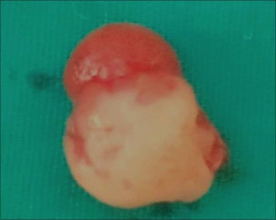
| Figure 1:Soft, cylindrical growth, 5 cm × 2 cm in dimension, pedunculated, situated near nasopalatine foramen
On histological examination, hyperkeratinized stratified squamous epithelium overlying dense connective tissue stroma was reported. The epithelium showed areas of keratin plugging along with collection of sebaceous glands in subepithelial regions. These lobules contained undifferentiated cells in the periphery and mature sebocytes with clear cytoplasm in the centre. Sheets, islands and cystic spaces of squamous epithelial cells and sebaceous cells were seen, without any cellular atypia or mitosis [Figure 2]. Underlying connective tissue was dense fibrous and collagenous containing numerous blood vessels along with areas of adipose tissue. Based on histological appearance, diagnosis of fibroepithelial hyperplasia with ectopic sebaceous gland and keratotic plugging was given.

| Figure 2:An area of keratotic plugging along with invaginating hyperplastic epithelium, collection of sebaceous glands in the subepithelial region with multiple lobules seen to be opening into a single duct
Discussion
SGH is rarely diagnosed intraorally. Till date, only 26 cases of SGH have been reported in the English language literature. Ectopic sebaceous glands are found in 80%–85% of adults in the buccal mucosa and lip and less often on the palate, gingivae, and tongue.[7] There are many theories regarding its etiology. One of these theories claimed that FEP develops secondary to a focal loss of elastic tissue. Another theory suggests that these polyps are a collection of several tissues that represents a hamartoma with a slow rate of growth or fibroma that exhibit the feature of a benign lesion.[8]
The mean age of occurrence is in the third decade of life with a slight female predilection. The present case was a 25-year-old male with a cylindrical polyp hanging from the anterior region of hard palate present since 3 years. SGH originates in the sebaceous follicles.[9]
Sebaceous gland lobules may arise from the inclusion of ectoderm in the oral cavity during the fusion of the maxillary and mandibular processes during the development of the embryo.[10] Microscopically, SGH originates from a single enlarged sebaceous gland composed of numerous lobules grouped from centrally located wide sebaceous duct. Similar histological picture was seen in the present case with keratin plugging of the ductal opening.
Sebaceous glands are composed of lobules containing a peripheral single or double layer of germinative flattened squamous cells, which proliferate and differentiate to the typical mature, clear sebaceous cells. The secretory product of sebaceous glands is called sebum.[11,12] Sebum comprises a mixture of lipids, including triglycerides, waxes, squalene, and cholesterol and its esters. Sebum may have weak antibacterial and antifungal properties and can be demonstrated using stain like Sudan Black B.[13]
Conclusion
The inflammatory hyperplastic lesions are difficult to diagnose since all lesions exhibit overlapping clinical features. Their presence may obstruct the insertion of an oral prosthesis, cause difficulty with mastication or speech, or even cause bleeding and ulceration following a secondary infection. Therefore these lesions must be surgically excised. The present article represents a rare case of fibroepithelial polyp showing sebaceous gland hyperplasia.
Financial support and sponsorship
Nil.
Conflicts of interest
There are no conflicts of interest.
References
- Halim DS, Pohchi A, Pang EE. The prevalence of fibroma in oral mucosa among patient attending USM dental clinic year 2006-2010. Indonesian J Dent Res 2010;1:61-6.
- Daley TD, Wysocki GP, Wysocki PD, Wysocki DM. The major epulides: Clinicopathological correlations. J Can Dent Assoc 1990;56:627-30.
- Prasanna J, Sehrawat S. Fibroepithelial hyperplasia: Rare, self-limiting condition – Two case reports. J Adv Oral Res 2011;2:63-9.
- Dent CD, Hunter WE, Svirsky JA. Sebaceous gland hyperplasia: Case report and literature review. J Oral Maxillofac Surg 1995;53:936-8.
- Daley T. Pathology of intraoral sebaceous glands. J Oral Pathol Med 1993;22:241-5.
- Daley TD. Intraoral sebaceous hyperplasia. Diagnostic criteria. Oral Surg Oral Med Oral Pathol 1993;75:343-7.
- Ferguson JW, Geary CP, MacAlister AD. Sebaceous cell adenoma. Rare intra-oral occurrence of a tumour which is a frequent marker of Torre's syndrome. Pathology 1987;19:204-8.
- Kumar A, Hasin N, Sinha AK, Kumar S, Bhadani P. Giant fibro epithelial polyp in a young girl: A rare case report. Intern J Surg Case Reports 2017;38:83-5.
- Lever WF, Schaumburg-Lever G. Histopathology of the Skin. Philadelphia, PA: Lippincott; 1975. p. 500.
- Shafer WF, Hine MK, Levy BM. A Textbook of Oral Pathology. 4th ed. Philadelpehia, PA: Saunders; 1983. p. 20.
- Kaminagakura E, Andrade CR, Rangel AL, Coletta RD, Graner E, Almeida OP, et al. Sebaceous adenoma of oral cavity: Report of case and comparative proliferation study with sebaceous gland hyperplasia and Fordyce's granules. Oral Dis 2003;9:323-7.
- Takeda Y, Satoh M, Nakamura S, Takamaru H. Sebaceous gland hyperplasia in an intraoral fibrous polyp. Pathol Int 2004;54:877-9.
- Somashekara KG, Lakshmi S, Priya NS. A rare case of sebaceous adenoma of the palate, with literature review. J Laryngology and Otology 2011;1-3.
| Figure 1:Soft, cylindrical growth, 5 cm × 2 cm in dimension, pedunculated, situated near nasopalatine foramen
| Figure 2:An area of keratotic plugging along with invaginating hyperplastic epithelium, collection of sebaceous glands in the subepithelial region with multiple lobules seen to be opening into a single duct
References
- Halim DS, Pohchi A, Pang EE. The prevalence of fibroma in oral mucosa among patient attending USM dental clinic year 2006-2010. Indonesian J Dent Res 2010;1:61-6.
- Daley TD, Wysocki GP, Wysocki PD, Wysocki DM. The major epulides: Clinicopathological correlations. J Can Dent Assoc 1990;56:627-30.
- Prasanna J, Sehrawat S. Fibroepithelial hyperplasia: Rare, self-limiting condition – Two case reports. J Adv Oral Res 2011;2:63-9.
- Dent CD, Hunter WE, Svirsky JA. Sebaceous gland hyperplasia: Case report and literature review. J Oral Maxillofac Surg 1995;53:936-8.
- Daley T. Pathology of intraoral sebaceous glands. J Oral Pathol Med 1993;22:241-5.
- Daley TD. Intraoral sebaceous hyperplasia. Diagnostic criteria. Oral Surg Oral Med Oral Pathol 1993;75:343-7.
- Ferguson JW, Geary CP, MacAlister AD. Sebaceous cell adenoma. Rare intra-oral occurrence of a tumour which is a frequent marker of Torre's syndrome. Pathology 1987;19:204-8.
- Kumar A, Hasin N, Sinha AK, Kumar S, Bhadani P. Giant fibro epithelial polyp in a young girl: A rare case report. Intern J Surg Case Reports 2017;38:83-5.
- Lever WF, Schaumburg-Lever G. Histopathology of the Skin. Philadelphia, PA: Lippincott; 1975. p. 500.
- Shafer WF, Hine MK, Levy BM. A Textbook of Oral Pathology. 4th ed. Philadelpehia, PA: Saunders; 1983. p. 20.
- Kaminagakura E, Andrade CR, Rangel AL, Coletta RD, Graner E, Almeida OP, et al. Sebaceous adenoma of oral cavity: Report of case and comparative proliferation study with sebaceous gland hyperplasia and Fordyce's granules. Oral Dis 2003;9:323-7.
- Takeda Y, Satoh M, Nakamura S, Takamaru H. Sebaceous gland hyperplasia in an intraoral fibrous polyp. Pathol Int 2004;54:877-9.
- Somashekara KG, Lakshmi S, Priya NS. A rare case of sebaceous adenoma of the palate, with literature review. J Laryngology and Otology 2011;1-3.