 PDF
PDF  Views
Views  Share
Share


Ovarian Endometriosis with Borderline Serous Tumor- Association or Coincidence – A Case Report and Review of Literature
CC BY-NC-ND 4.0 · Indian J Med Paediatr Oncol 2020; 41(03): 406-408
DOI: DOI: 10.4103/ijmpo.ijmpo_209_18
Abstract
Endometriosis is the presence of endometrial glands outside the endometrium, and ovary is the common site for endometriosis. Endometriosis can also transform into malignant tumors. When endometriosis is present within the tumors, the term endometriosis-derived tumor applies, whereas when endometriosis is recognized adjacent to the tumor, it is called as endometriosis-associated tumor. Borderline serous tumor is surface epithelial ovarian tumor. The endometriosis-associated ovarian malignancies are clear cell adenocarcinoma and endometrioid adenocarcinoma, whereas serous and mucinous are rare malignancies with endometriosis. Here, we are presenting a case report in which endometriosis was associated with borderline serous tumor.
Publication History
Received: 20 September 2018
Accepted: 10 November 2019
Article published online:
28 June 2021
© 2020. Indian Society of Medical and Paediatric Oncology. This is an open access article published by Thieme under the terms of the Creative Commons Attribution-NonDerivative-NonCommercial-License, permitting copying and reproduction so long as the original work is given appropriate credit. Contents may not be used for commercial purposes, or adapted, remixed, transformed or built upon. (https://creativecommons.org/licenses/by-nc-nd/4.0/.)
Thieme Medical and Scientific Publishers Pvt. Ltd.
A-12, 2nd Floor, Sector 2, Noida-201301 UP, India
Abstract
Endometriosis is the presence of endometrial glands outside the endometrium, and ovary is the common site for endometriosis. Endometriosis can also transform into malignant tumors. When endometriosis is present within the tumors, the term endometriosis-derived tumor applies, whereas when endometriosis is recognized adjacent to the tumor, it is called as endometriosis-associated tumor. Borderline serous tumor is surface epithelial ovarian tumor. The endometriosis-associated ovarian malignancies are clear cell adenocarcinoma and endometrioid adenocarcinoma, whereas serous and mucinous are rare malignancies with endometriosis. Here, we are presenting a case report in which endometriosis was associated with borderline serous tumor.
Introduction
The ovary is a common site for endometriosis. Endometriosis is the presence of endometrium glands and stroma outside the uterus.[1] It is well recognized that malignant transformation can occurs in endometriosis. Nishida et al. reported 18 cases of atypical endometriosis and one case of ovarian carcinoma in 147 cases of ovarian endometriosis, thus the incidence of malignancy in ovarian endometriosis is 0.7%.[2] However, among the malignancies, clear cell adenocarcinoma and endometrioid adenocarcinoma are the common malignancies associated with endometriosis. Serous and mucinous tumors are infrequently seen with endometriosis.[3] Here, we present a case report of a 40-year-old female who clinically presented with abnormal uterine bleeding (AUB) and ovarian mass. On histopathological examination, it was diagnosed as a borderline serous tumor with endometriosis.
Case Report
A 40-year-old female presented to the outpatient setting with AUB and abdominal mass. On ultrasonography examination, it was diagnosed as an ovarian cyst. Hysterectomy with bilateral salpingo-oophorectomy was performed, and the specimen was sent in 10%-formal saline for histopathological analysis. Grossly, hysterectomy with bilateral salpingo-oophorectomy measured 12 cm × 9 cm × 6 cm, on further sectioning, multiple leiomyomas measuring 1 to 1.5 cm in size were also seen in the myometrium, [Figure 1] whereas, the left sided ovary showed hemorrhagic luteal cyst and right sided ovary measured 8 cm × 6 cm × 4 cm, which on cutting was uniloculated, with papillary architecture and filled with serous fluid [Figure 2]. On histopathology examination, right sided ovary showed a borderline serous tumor, and associated endometriosis [Figure 3] and [Figure 4].{Figure 1}{Figure 2}{Figure 3}{Figure 4}
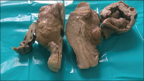
| Figure 1:Gross examination of hysterectomy showing subserosal and intra mural-leiomyoma, whereas right-sided ovary having borderline serous cystadenoma with endometriosis

| Figure.2:Gross picture of right-sided ovary showing borderline serous cystadenoma with endometriosis
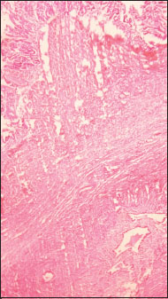
| Figure.3:Borderline serous cystadenoma with benign endometriosis, H and E (×40)
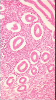
| Figure.4:Endometrial glands and its stroma lying in ovarian stroma, H and E (×100)
Discussion
Ovarian tumors are common tumors of the female genital tract. The WHO classification of ovarian tumor includes surface epithelial tumor, germ cell tumor, sex cord stromal tumour, miscellaneous and metastatic tumors.[4],[5] Surface epithelial tumor constitutes 60%–70%-of all ovarian neoplasms and 90%-of malignant ovarian tumors. The common epithelial tumors are serous, mucinous, and endometrioid. Serous tumors are divided into benign, borderline, and malignant.[5] Endometriosis is defined as heterotopic presence of endometrium, that is, endometrial glands with its stroma outside the endometrium. Exact pathology is not known, but some theories believe that reflux of endometrial tissue through fallopian tube at the time of menstruation, coelomic metaplasia, embryonic cell rest lymphatic and vascular dissemination have been proposed to explain the theory of endometriosis.[6] The pathogenesis is multifactorial including genetic, hormonal, environment, and role of immune system.[7] The ovary is the most common site of endometriosis followed by pelvic structures. In 1925, for the first time, Sampson explained the association between endometriosis and ovarian carcinoma.[8] Stern et al. reported that ovarian malignancies were associated with endometriosis in 3.2%–10%-of cases.[9] Histologically, in majority of the patients with ovarian epithelial tumors and associated endometriosis, the ovarian tumor is of endometrioid (53%) or clear cell lineage (22%); and in most cases with associated endometriosis serous or mucinous ovarian epithelial tumors, it was more frequently borderline, which also justifies the better prognosis of ovarian epithelial tumors associated with endometriosis.[10] The present case also confirms the borderline nature of serous ovarian tumor with associated endometriosis.
The significance of endometriosis arises because of its malignant potential, pathology might be high estrogen concentration which leads to malignant proliferation of endometriotic cyst or due to mutation in the AR1D1A gene and consequent loss of BAF250a expression, similarly, iron produced in endometriotic cyst promotes oxidative stress, which causes genetic mutation and malignant progression of ovarian cyst.[11]
Conclusion
It is uncommon to find an association of borderline serous tumor with endometriosis, because the incidence is higher with clear cell and endometrioid carcinoma, but still possibilities are always there. Thus, histopathological evaluation of every ovarian mass must be carried out to determine the ovarian tumor along with the endometriosis, especially to analyze the malignant potential of endometriosis.
Conflict of Interest
There are no conflicts of interest.
References
- Rosai J, Sarin YK. editor. Uterus-Corpus. In: Text Book of Rosai Ackerman's Surgical Pathology. St Louis, Missouri: Mosby Elsevier 2004; 9: 1577
- Nishida M, Watanabe K, Sato N, Ichikawa Y. Malignant transformation of ovarian endometriosis. Gynecol Obstet Invest 2000; 50 Suppl 1: 18-25
- Valenzuela P, Ramos P, Redondo S, Cabrera I, Ruiz A. Endometrioid adenocarcinoma of the ovary and endometriosis. Eur J Obstet Gynecol Reprod Biol 2007; 134: 83-6
- Christopher CP. The female genital tract. In: Kumar V, Abbas AK, Fausto N. editors Robbins and Cotran Pathologic basis of disease. New Delhi: Saunders Elsevier; 2007. 7. 1093
- Mohan H. editors The female genital tract. In: Textbook of Pathology. New Delhi: Jaypee Brothers Medical Publishers; 2010. 6. 721-53
- Worley MJ, Welch WR, Berkowitz RS, Ng SW. Endometriosis-associated ovarian cancer: A review of pathogenesis. Int J Mol Sci 2013; 14: 5367-79
- Giudice LC, Kao LC. Endometriosis. Lancet 2004; 364: 1789-99
- trong> Akbarzadeh-Jahromi SM, Shekarkhar G, Sari AslaniF, Azarpira N, Heidari EsfahaniM, Momtahan M. et al. Prevalence of endometriosis in malignant epithelial ovarian tumor. Arch Iran Med 2015; 18: 844-8
- Stern RC, Dash R, Bentley RC, Snyder MJ, Haney AF, Robboy SJ. Malignancy in endometriosis: Frequency and comparison of ovarian and extraovarian types. Int J Gynecol Pathol 2001; 20: 133-9
- Bas-Esteve E, Pérez-Arguedas M, Guarda-Muratori GA, Acién M, Acién P. Endometriosis and ovarian cancer: Their association and relationship. Eur J Obstet Gynecol Reprod Biolx 2019; 3: 100053
- Moris Brilhante AV, Augusto KL, Portela MC, Gabriele Sucupira LC, Freistas Oliveira LA, Verissimo Pouchaim AJ. et al. Endometriosis and ovarian cancer: An integrative review (Endometriosis and ovarian cancer) . Asian Pac J Cancer Prev 2017; 18: 11-6
Address for correspondence
Publication History
Received: 20 September 2018
Accepted: 10 November 2019
Article published
online:
28 June 2021
© 2020. Indian Society of Medical and Paediatric Oncology. This is an open access article published by Thieme under the terms of the Creative Commons Attribution-NonDerivative-NonCommercial-License, permitting copying and reproduction so long as the original work is given appropriate credit. Contents may not be used for commercial purposes, or adapted, remixed, transformed or built upon. (https://creativecommons.org/licenses/by-nc-nd/4.0/.)
Thieme Medical and Scientific Publishers Pvt. Ltd.
A-12, 2nd Floor, Sector 2,
Noida-201301 UP, India
| Figure 1:Gross examination of hysterectomy showing subserosal and intra mural-leiomyoma, whereas right-sided ovary having borderline serous cystadenoma with endometriosis
| Figure.2:Gross picture of right-sided ovary showing borderline serous cystadenoma with endometriosis
| Figure.3:Borderline serous cystadenoma with benign endometriosis, H and E (×40)
| Figure.4:Endometrial glands and its stroma lying in ovarian stroma, H and E (×100)
References
- Rosai J, Sarin YK. editor. Uterus-Corpus. In: Text Book of Rosai Ackerman's Surgical Pathology. St Louis, Missouri: Mosby Elsevier 2004; 9: 1577
- Nishida M, Watanabe K, Sato N, Ichikawa Y. Malignant transformation of ovarian endometriosis. Gynecol Obstet Invest 2000; 50 Suppl 1: 18-25
- Valenzuela P, Ramos P, Redondo S, Cabrera I, Ruiz A. Endometrioid adenocarcinoma of the ovary and endometriosis. Eur J Obstet Gynecol Reprod Biol 2007; 134: 83-6
- Christopher CP. The female genital tract. In: Kumar V, Abbas AK, Fausto N. editors Robbins and Cotran Pathologic basis of disease. New Delhi: Saunders Elsevier; 2007. 7. 1093
- Mohan H. editors The female genital tract. In: Textbook of Pathology. New Delhi: Jaypee Brothers Medical Publishers; 2010. 6. 721-53
- Worley MJ, Welch WR, Berkowitz RS, Ng SW. Endometriosis-associated ovarian cancer: A review of pathogenesis. Int J Mol Sci 2013; 14: 5367-79
- Giudice LC, Kao LC. Endometriosis. Lancet 2004; 364: 1789-99
- trong> Akbarzadeh-Jahromi SM, Shekarkhar G, Sari AslaniF, Azarpira N, Heidari EsfahaniM, Momtahan M. et al. Prevalence of endometriosis in malignant epithelial ovarian tumor. Arch Iran Med 2015; 18: 844-8
- Stern RC, Dash R, Bentley RC, Snyder MJ, Haney AF, Robboy SJ. Malignancy in endometriosis: Frequency and comparison of ovarian and extraovarian types. Int J Gynecol Pathol 2001; 20: 133-9
- Bas-Esteve E, Pérez-Arguedas M, Guarda-Muratori GA, Acién M, Acién P. Endometriosis and ovarian cancer: Their association and relationship. Eur J Obstet Gynecol Reprod Biolx 2019; 3: 100053
- Moris Brilhante AV, Augusto KL, Portela MC, Gabriele Sucupira LC, Freistas Oliveira LA, Verissimo Pouchaim AJ. et al. Endometriosis and ovarian cancer: An integrative review (Endometriosis and ovarian cancer) . Asian Pac J Cancer Prev 2017; 18: 11-6