 PDF
PDF  Views
Views  Share
Share


Radiofrequency Ablation of Osteoid Osteoma in Common and Technically Challenging Locations in Pediatric Population
CC BY-NC-ND 4.0 · Indian J Med Paediatr Oncol 2017; 38(03): 302-305
DOI: DOI: 10.4103/ijmpo.ijmpo_61_16
|
Publication History
Article published online:
04 July 2021
© 2017. Indian Society of Medical and Paediatric Oncology. This is an open access article published by Thieme under the terms of the Creative Commons Attribution-NonDerivative-NonCommercial-License, permitting copying and reproduction so long as the original work is given appropriate credit. Contents may not be used for commercial purposes, or adapted, remixed, transformed or built upon. (https://creativecommons.org/licenses/by-nc-nd/4.0/.)
Thieme Medical and Scientific Publishers Pvt. Ltd.
A-12, 2nd Floor, Sector 2, Noida-201301 UP, India
Abstract
Context:
Percutaneous radiofrequency ablation (RFA) of osteoid osteoma has a high technical and clinical success rate. However, there is limited data on its use in the pediatric population, especially in technically challenging locations.
Objective:
The objective of this study was to assess the safety and efficacy of CT-guided percutaneous RFA of osteoid osteoma in pediatric population.
Subjects and Methods:
From June 2009 to May 2014, thirty patients with osteoid osteoma were treated with CT-guided RFA in common (25 cases) and technically challenging (five cases: four near articular surface and one in sacrum) locations. Therapy was performed under general anesthesia with a three-array expandable RF probe for 6 min at 90°C and power of 60–100 W. The patients were discharged next day under instruction. The treatment success was evaluated in terms of pain relief before and after (1 day, 1 month, and 6 months) treatment.
Results:
Technical success was achieved in all patients (100%). Primary clinical success was 96.66% (29 of total 30 patients) despite the pediatric population and atypical location. One patient had persistent pain after 1 month duration and were treated successfully with a second procedure (secondary success rate was 100%). One patient had immediate complication of weakness of right hand and fingers extension. No delayed complications were observed.
Conclusions:
CT-guided RFA is relatively safe and highly effective for treatment of osteoid osteoma in pediatric population, even in technically difficult locations.
Advance in Knowledge:
Our study showed that if technical success is 100% and if strict desired temperature (90°C) can be maintained for desired time (6 min) using controlled power (wattage) delivery (60–100 W), then high clinical success can be achieved even in pediatric population similar to adult population.
Introduction
Osteoid osteoma is a benign bone tumor consisting of an osteoid nidus in a highly vascular connective tissue stroma. These tumors are exquisitely painful and demonstrate characteristic findings on clinical and radiographic examinations.[1,2] The traditional treatment of osteoid osteoma consists of surgical en bloc excision.[2,3,4] The major drawback of this traditional approach is prolonged surgery and hospital stay, weakening of the bone requiring a prophylactic fixation and its subsequent removal, and additional time off school due to open surgery. To overcome this drawback, different minimally invasive techniques have been described as alternative therapeutic options (radiofrequency ablation [RFA], laser photocoagulation, and percutaneous resection).[3,4,5,6,7] CT guidance has become a very useful and easy method for the percutaneous treatment of these lesions.[8] We started using CT-guided RFA for osteoid osteoma 5 years ago and immediately noted many beneficial effects in terms of duration of hospital stay, morbidity, and overall patient comfort prompting us to do this study. We report our experience with CT-guided percutaneous RFA of osteoid osteoma in common and technically challenging locations in 30 pediatric patients and evaluate technical and clinical results.
Subjects and Methods
This is a retrospective observational study of 30 pediatric and adolescent patients who underwent CT-guided percutaneous RFA of osteoid osteoma between June 2009 and May 2014. All patients reported severe pain that usually increased at night and required nonsterioidal anti-inflammatory drugs for pain relief. The osteoid osteoma was diagnosed from clinical and imaging findings (radiography, CT, and/or magnetic resonance imaging scan), demonstrating a nidus and other findings that are typical of osteoid osteoma. Patients and their legal guardians were fully informed of the procedure and of the surgical and medical alternatives, and informed consent was obtained.
All procedures were performed under general anesthesia in the CT room on six-slice CT scanner (Philips Brilliance, Massachusetts, USA). Grounding pads were placed. Under aseptic precautions, a guidewire (K-wire) was drilled into the center of the nidus using either a hand drill (Aesculap Inc. B Braun, Center Valley, PA, USA) or a battery operated drill (Stryker Corp., Kalamazoo, MI, USA) with a cannulated drill bit (2.5–4.5 mm) (Zimmer, Warsaw, IN, USA) along the planned tract by the pediatric orthopedist; subsequently, a 4 mm cannulated bone drill was exchanged for a 5F sheath over an Amplatz wire. RFA needle (RITA SDE StarBurst Probe [17-gauge 2 cm diameter, 12 cm long, three tines] Angiodynamics, Inc., GA, USA) was inserted into the nidus of osteoid osteoma through the sheath. The tines were opened and confirmed to be within the lesion with check CT sections. Using the radiofrequency waves from RF generator (Model 1500X; Angiodynamics, Inc., GA, USA), the tines were heated to a target temperature of 90°C and power of 60–100 W and the peak temperature was maintained for 6 min. After 6 min, the tines were withdrawn into the probe, and then, the RF probe removed. Next day, the patient was clinically evaluated for type and severity of pain.
The treatment success was evaluated in terms of pain relief before and after procedure (1 day, 1 month and 3 months). Patients were also contacted by telephone at 6 months for follow-up regarding complications or recurrence of pain. Technical success was defined as the ability to localize the radiolucent nidus and placement of an electrode under CT guidance with ablation performed for the desired period. Clinical success was defined as complete relief of pain without the use of oral pain medication within 1 month of the procedure.
Results
From June 2009 to May 2014, thirty pediatric patients underwent CT-guided RF ablation of osteoid osteoma. There were 25 boys and 5 girls with male to female ratio of 5:1. Their age ranged between 4 and 20 years with a mean age of 13.16 years. Lesions were grouped into common and challenging location. Among the common location (n = 25), lesions were located in the femur (n = 21) [Figure 1] and tibia (n = 4). Among the challenging locations (n = 5), four were near articular surface (one each at glenoid fossa of right scapula, head of right radius, talocalcalcaneal joint of right calcaneum [Figure 2], and left femoral head) and one was in left sacrum.
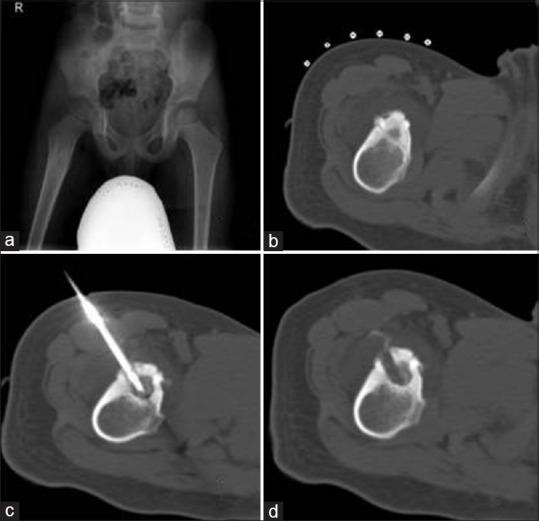
| Figure 1:An 11-year-old boy with a right proximal femur osteoid osteoma. Radiography (a) and computed tomography scan bone window axial image (b) radiolucent nidus and surrounding reactive sclerosis. (c) Computed tomography scan shows radiofrequency ablation probe in situ in nidus. (d) Computed tomography scan postprocedure shows radiofrequency ablation tract

| Figure 2:A 17-year-old boy with a right calcaneal osteoid osteoma near the talocalcaneal joint margin. (a) Magnetic resonance imaging (short tau inversion recovery) sagittal image shows central nidus surrounded by hypointense reactive sclerosis. Rest of the surrounding calcaneum appear hyperintense due to reactive edema due to osteoid osteoma. (b) Computed tomography scan shows radiofrequency ablation probe in situ in nidus
Technical success was achieved in all patients (100%). The number of ablations per treatment session was one. The time of ablation was 6 min for each setting. The duration of the procedures ranged from 60 to 150 min. Primary clinical success was 96.66% (29 of total 30 patients) despite pediatric population and challenging location. One patient with an osteoid osteoma in the shaft of right femur had persistent pain after 1 month and was treated successfully with a second procedure (secondary success rate 100%). One patient from the challenging location group with osteoid osteoma at right radial head had immediate postprocedure weakness of wrist and finger extension as the lesion was very close to posterior interosseous nerve which slowly recovered with physiotherapy. No delayed complications were observed.
Discussion
Since the promising results of Rosenthal et al.[9] in the management of osteoid osteoma with RF ablation, a large number of studies evaluating RF ablation of osteoid osteoma have been reported in the literature. Most of these studies found very high technical success rates (100%) and good primary success rates with a single session of ablation ranging from 76% to 100%.[10,11,12,13,14,15,16,17] The secondary success rate after repeated ablations ranged from 87% to 100%. Hence, this minimally invasive technique has become the method of choice for treatment of osteoid osteomas, provided that the diagnosis is based on a typical clinical, scintigraphic, and CT presentation.[18] To our knowledge, there are few reports in the literature regarding the role of percutaneous RF ablation in treating osteoid osteomas in children at atypical locations.
Our study of RFA of osteoid osteoma included 30 pediatric patients. Our technical success rate was 100%, which is similar to most of the other studies (100%). In our study, in pediatric population, the primary and secondary clinical success ratings were 96.66% and 100%, respectively, which are comparable to success rates in most other studies in adults where primary success and secondary success ranged 76%–100% and 87%–100%, respectively.[10,11,12,13,14,15,16,17] In a study done by Donkol et al. on efficacy of RFA of osteoid osteoma in children[19] showed that the technical success, primary clinical success, and secondary clinical success rates were 91.3%, 78.2%, and 82.6%, respectively. Donkol et al.[19] and Vanderschueren et al.[20] in their studies showed that lower age can be a risk factor for lower clinical success rate. This can be explained by the greater technical difficulty during ablation of osteoid osteoma in children due to the small body mass, difficulty in positioning and fixation of the needle, and shorter ablation time (range 2–6 min in their study) for each procedure. Our study showed that if technical success is 100% and if strict desired temperature (90°C) can be maintained for desired time (6 min) using controlled power (wattage) delivery (60–100 W), then high clinical success can be achieved even in pediatric population similar to adult population.
In our study, transient radial nerve palsy was the only complication. This happened in a child with radial head (technically challenging position) osteoid osteoma who had developed an immediate postprocedure weakness of wrist and finger extension as the lesion was very close to posterior interosseous nerve; this slowly recovered with physiotherapy. There are few case reports of likely articular cartilage damage in weight-bearing joints such as acetabulum following CT-guided percutaneous RF ablation of juxta-articular osteoid osteoma, but it was not confirmed whether the articular cartilage was damaged only by head due to RFA or if it had also been weakened before by the osteoid osteoma.[21] Another study by Papagelopoulos et al.[22] on RFA of intra-articular osteoid osteoma of the hip showed that there is a good ossification and bone regeneration following RF ablation and it is safe and effective treatment even for intra-articular lesions. No delayed complications were observed in our study.
In comparison to percutaneous CT-guided curettage of osteoid osteoma, during RF ablation, there is a lack of confirmation of histological diagnosis. However, there are other disadvantages of curettage technique that it is little more traumatic with complications of neuropraxia, skin abrasions, damage to blood vessels, especially in femur leading to avascular necrosis and frequently incomplete curettage.[23]
Limitations of our study are that it is an observational study, lack of confirmation of histological diagnosis, and lack of imaging follow-up.
Conclusions
Percutaneous CT-guided RF ablation is a relatively safe, highly effective, and minimally invasive procedure for the treatment of osteoid osteoma in pediatric population despite atypical location.
Financial support and sponsorship
Nil.
Conflicts of interest
There are no conflicts of interest.
References
- Frosch MP, Anthony DC, de Girolami U. The central nervous system. In: Kumar V, Abbas AK, Fausto N, Aster JC, editors. Robbins Cotran Pathologic Basis of Disease. 8th ed. Philadelphia: Saunders Elsevier; 2010.
- Koss LG, Rodriguez CA. The central nervous system. In: Koss LG, Melamed MR. Koss's diagnostic cytology and its histopathologic bases. 5th ed. Vol. 2. Philadelphia: Lippincott Williams and Wilkins; 2006. p. 1523-43.
- Adesina AM. Intraoperative consultation in the diagnosis of pediatric brain tumors. Arch Pathol Lab Med 2005;129:1653-60.
- Brainard JA, Prayson RA, Barnett GH. Frozen section evaluation of stereotactic brain biopsies: Diagnostic yield at the stereotactic target position in 188 cases. Arch Pathol Lab Med 1997;121:481-4.
- Brommeland T, Lindal S, Straume B, Dahl IL, Hennig R. Does imprint cytology of brain tumours improve intraoperative diagnoses? Acta Neurol Scand 2003;108:153-6.
- Burger PC, Nelson JS. Stereotactic brain biopsies: Specimen preparation and evaluation. Arch Pathol Lab Med 1997;121:477-80.
- Burger PC. Smears and Frozen Sections in Surgical Neuropathology. Baltimore: PB Medical Publishing; 2009.p.3-10, 163-287, 335-47, 359-99.
- Firlik KS, Martinez AJ, Lunsford LD. Use of cytological preparations for the intraoperative diagnosis of stereotactically obtained brain biopsies: A 19-year experience and survey of neuropathologists. J Neurosurg 1999;91:454-8.
- Folkerth RD. Smears and frozen sections in the intraoperative diagnosis of central nervous system lesions. Neurosurg Clin N Am 1994;5:1-18.
- Hayden R, Cajulis RS, Frias-Hidvegi D, Brody BA, Yu G, Levy R. Intraoperative diagnostic techniques for stereotactic brain biopsy: Cytology versus frozen-section histopathology. Stereotact Funct Neurosurg 1995;65:187-93.
- Joseph JT. Diagnostic Neuropathology Smears. Philadelphia: Lippincott Williams and Wilkins; 2007. p. 1-234.
- Olasode BJ, Ironside JW. The brain smear, a rapid affordable intraoperative diagnostic technique for brain tumours appropriate for Africa. Trop Doct 2004;34:223-5.
- Mitra S, Kumar M, Sharma V, Mukhopadhyay D. Squash preparation: A reliable diagnostic tool in the intraoperative diagnosis of central nervous system tumors. J Cytol 2010;27:81-5.
- Cahill EM, Hidvegi DF. Crush preparations of lesions of the central nervous system. A useful adjunct to the frozen section. Acta Cytol 1985;29:279-85.
- Powell SZ. Intraoperative consultation, cytologic preparations, and frozen section in the central nervous system. Arch Pathol Lab Med 2005;129:1635-52.
| Figure 1:An 11-year-old boy with a right proximal femur osteoid osteoma. Radiography (a) and computed tomography scan bone window axial image (b) radiolucent nidus and surrounding reactive sclerosis. (c) Computed tomography scan shows radiofrequency ablation probe in situ in nidus. (d) Computed tomography scan postprocedure shows radiofrequency ablation tract
| Figure 2:A 17-year-old boy with a right calcaneal osteoid osteoma near the talocalcaneal joint margin. (a) Magnetic resonance imaging (short tau inversion recovery) sagittal image shows central nidus surrounded by hypointense reactive sclerosis. Rest of the surrounding calcaneum appear hyperintense due to reactive edema due to osteoid osteoma. (b) Computed tomography scan shows radiofrequency ablation probe in situ in nidus
References
- Frosch MP, Anthony DC, de Girolami U. The central nervous system. In: Kumar V, Abbas AK, Fausto N, Aster JC, editors. Robbins Cotran Pathologic Basis of Disease. 8th ed. Philadelphia: Saunders Elsevier; 2010.
- Koss LG, Rodriguez CA. The central nervous system. In: Koss LG, Melamed MR. Koss's diagnostic cytology and its histopathologic bases. 5th ed. Vol. 2. Philadelphia: Lippincott Williams and Wilkins; 2006. p. 1523-43.
- Adesina AM. Intraoperative consultation in the diagnosis of pediatric brain tumors. Arch Pathol Lab Med 2005;129:1653-60.
- Brainard JA, Prayson RA, Barnett GH. Frozen section evaluation of stereotactic brain biopsies: Diagnostic yield at the stereotactic target position in 188 cases. Arch Pathol Lab Med 1997;121:481-4.
- Brommeland T, Lindal S, Straume B, Dahl IL, Hennig R. Does imprint cytology of brain tumours improve intraoperative diagnoses? Acta Neurol Scand 2003;108:153-6.
- Burger PC, Nelson JS. Stereotactic brain biopsies: Specimen preparation and evaluation. Arch Pathol Lab Med 1997;121:477-80.
- Burger PC. Smears and Frozen Sections in Surgical Neuropathology. Baltimore: PB Medical Publishing; 2009.p.3-10, 163-287, 335-47, 359-99.
- Firlik KS, Martinez AJ, Lunsford LD. Use of cytological preparations for the intraoperative diagnosis of stereotactically obtained brain biopsies: A 19-year experience and survey of neuropathologists. J Neurosurg 1999;91:454-8.
- Folkerth RD. Smears and frozen sections in the intraoperative diagnosis of central nervous system lesions. Neurosurg Clin N Am 1994;5:1-18.
- Hayden R, Cajulis RS, Frias-Hidvegi D, Brody BA, Yu G, Levy R. Intraoperative diagnostic techniques for stereotactic brain biopsy: Cytology versus frozen-section histopathology. Stereotact Funct Neurosurg 1995;65:187-93.
- Joseph JT. Diagnostic Neuropathology Smears. Philadelphia: Lippincott Williams and Wilkins; 2007. p. 1-234.
- Olasode BJ, Ironside JW. The brain smear, a rapid affordable intraoperative diagnostic technique for brain tumours appropriate for Africa. Trop Doct 2004;34:223-5.
- Mitra S, Kumar M, Sharma V, Mukhopadhyay D. Squash preparation: A reliable diagnostic tool in the intraoperative diagnosis of central nervous system tumors. J Cytol 2010;27:81-5.
- Cahill EM, Hidvegi DF. Crush preparations of lesions of the central nervous system. A useful adjunct to the frozen section. Acta Cytol 1985;29:279-85.
- Powell SZ. Intraoperative consultation, cytologic preparations, and frozen section in the central nervous system. Arch Pathol Lab Med 2005;129:1635-52.