 PDF
PDF  Views
Views  Share
Share


Epidemiology of musculoskeletal tumors in Shiraz, south of Iran
CC BY-NC-ND 4.0 · Indian J Med Paediatr Oncol 2011; 32(04): 187-191
DOI: DOI: 10.4103/0971-5851.95138
Abstract
Background: Musculoskeletal tumors are rare, but their descriptive data in any region are important to reduce mortality rate and improve their management. Materials and Methods: Retrospectively, 426 pathologic reports from 1997 to 2008 were reviewed in Shiraz University Orthopedic Hospitals which are the main referral centers for musculoskeletal tumors in south of Iran. We collected and analyzed data on age, gender, anatomical site, and histopathologic types of musculoskeletal tumors. Results: Of the 426 cases, 60.1% were men and 39.9% were women. The commonest malignant bone tumors were osteosarcoma (89; 50.6%), metastasis (30; 17.0%), Ewing′s sarcoma (28; 15.9%), and chondrosarcoma (14; 8.0%). The most frequent benign bone tumors were osteochondroma (136; 63.9%), enchondroma (23; 10.8%), giant cell tumor (21; 9.9%), and osteoid osteoma (14; 6.6%). The femur was the most commonly involved site in musculoskeletal tumors. It was followed by the tibia in benign tumors and the humerus in malignant ones. Metastasis (28; 32.6%), soft tissue tumors (18; 20.9%), osteochondroma (10; 11.6%), and osteosarcoma (9; 10.5%) were the most diagnosed bone lesions in patients older than 40. Conclusion: There are no significant changes in epidemiology of musculoskeletal tumors in Shiraz, south of Iran, in comparison with other parts of the world.
Publication History
Article published online:
06 August 2021
© 2011. Indian Society of Medical and Paediatric Oncology. This is an open access article published by Thieme under the terms of the Creative Commons Attribution-NonDerivative-NonCommercial-License, permitting copying and reproduction so long as the original work is given appropriate credit. Contents may not be used for commercial purposes, or adapted, remixed, transformed or built upon. (https://creativecommons.org/licenses/by-nc-nd/4.0/.)
Thieme Medical and Scientific Publishers Pvt. Ltd.
A-12, 2nd Floor, Sector 2, Noida-201301 UP, India
Abstract
Background:
Musculoskeletal tumors are rare, but their descriptive data in any region are important to reduce mortality rate and improve their management.
Materials and Methods:
Retrospectively, 426 pathologic reports from 1997 to 2008 were reviewed in Shiraz University Orthopedic Hospitals which are the main referral centers for musculoskeletal tumors in south of Iran. We collected and analyzed data on age, gender, anatomical site, and histopathologic types of musculoskeletal tumors.
Results:
Of the 426 cases, 60.1% were men and 39.9% were women. The commonest malignant bone tumors were osteosarcoma (89; 50.6%), metastasis (30; 17.0%), Ewing's sarcoma (28; 15.9%), and chondrosarcoma (14; 8.0%). The most frequent benign bone tumors were osteochondroma (136; 63.9%), enchondroma (23; 10.8%), giant cell tumor (21; 9.9%), and osteoid osteoma (14; 6.6%). The femur was the most commonly involved site in musculoskeletal tumors. It was followed by the tibia in benign tumors and the humerus in malignant ones. Metastasis (28; 32.6%), soft tissue tumors (18; 20.9%), osteochondroma (10; 11.6%), and osteosarcoma (9; 10.5%) were the most diagnosed bone lesions in patients older than 40.
Conclusion:
There are no significant changes in epidemiology of musculoskeletal tumors in Shiraz, south of Iran, in comparison with other parts of the world.
INTRODUCTION
Malignant neoplasms are one of the main causes of mortality in the world. Skeletal system involvement as metastasis is common. The primary source comes usually from breast, prostate, kidney, lung, and thyroid.[1,2] Diagnosis of these lesions often changes the treatment plan.
Primary bone tumors are rare. They account for 0.2–0.5% of all malignancies in all ages[3] and comprise 3–5% of tumors diagnosed in European children below 15 years and 7–8% in adolescents between 15 and 19 years of age.[4] Incidence of primary malignant bone tumors is about 9 in 1 million people in a year. It is slightly higher in males than females (10/million/year vs. 8/million/year).[5] It is usual for these tumors to be diagnosed late because these neoplasms are uncommon; moreover, their presentations are vague with unspecific signs and symptoms. Sometimes they are recognized and treated like osteomyelitis or simple fracture before exact diagnosis. Therefore, basic epidemiology in each region can help doctors to diagnose and manage them earlier. Also, these studies can guide researchers to find particular risk factors in that area.
According to previous researches, distribution of primary bone tumors is variable in different parts of the world. Highest rates are seen in Europe and the USA, but Asian countries have lower incidence and prevalence.[4,6,7] Although there are several epidemiologic studies of musculoskeletal tumors in the neighboring countries of Iran,[8–11] to the best of our knowledge, this report is the first documented one in Iran.
MATERIALS AND METHODS
We reviewed retrospectively charts and pathologic reports of patients who had undergone musculoskeletal open or excisional biopsies from January 1997 to December 2008. Biopsies had been taken in Chamran and Namazi Hospitals of Shiraz University of Medical Sciences, two main referral centers in the south of Iran. Pathologic slides had been reported by three expert pathologists in musculoskeletal diseases. Also, final diagnosis had been correlated with clinical presentation and radiograph findings by an orthopedic tumor surgeon (senior author). It should be mentioned that although diagnosis of several bone tumors like osteoid osteoma, osteochondroma, and nonossifying fibroma was made clinically and radiographically, open biopsies had been taken due to equivocal diagnosis. Moreover, some reports were the result of excisional biopsies (like osteochondroma and osteoid osteoma). We collected data on age, sex, anatomical site, and histopathologic type of 426 patients.
On the basis of pathologic reports, the tumors were classified as benign bone tumors, malignant bone tumors, and soft tissue tumors. The latter comprised malignant ones like rhabdomyosarcoma, fibrosarcoma, etc. Benign soft tissue tumors were excluded. There were some nonspecific sarcomas like small cell malignant sarcoma and high-grade malignant sarcoma in pathologic reports. We classified them as others in the results.
Description statistics were performed using SPSS software version 18.0 for windows (SPSS Inc., Chicago, IL, USA). Frequency and percentage of the mentioned variables were calculated.
RESULTS
Totally, 426 cases were analyzed. Male to female ratio was 1.5. There were 170 women (39.9%) and 256 men (60.1%). The oldest patient was an 84-year-old man with metastasis. There were two 4-year-old children as the youngest patients, with pathologic reports of osteofibrous dysplasia and soft tissue tumor. As shown in Table 1, 176 patients had malignant bone tumors with the peak frequency seen in patients of age between 5 and 25 years. Also, large proportion of benign bone tumors (161; 75.5%) was seen in patients younger than 25 years of age. Soft tissue tumors occurred more (10; 27.1%) in the age group of 45–54 years.
Table 1
Distribution of musculoskeletal tumors by age
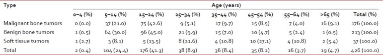
| Overall, the most common pathologic types of bone tumors were osteochondroma (136; 35.0%) and osteosarcoma (89; 22.9%). The femur (159; 40.9%) was the most common anatomical site of bone tumors, followed by the tibia (84; 21.6%), and the humerus (52; 13.4%).
References
- Coleman RE. Clinical features of metastatic bone disease and risk of skeletal morbidity. Clin Cancer Res 2006;12:6243s-9s.
- Lewis VO. What′s new in musculoskeletal oncology. J Bone Joint Surg Am 2007;89:1399-407.
- Fletcher CD, Unni KK, Mertens F, editors. Pathology and genetics of tumours of soft tissue and bone. Vol. 5 of World Health Organization classification of tumours. Lyon, France: IARC Press, 2002.
- Stiller CA, Bielack SS, Jundt G, Steliarova-Foucher E. Bone tumours in European children and adolescents, 1978-1997. Report from the Automated Childhood Cancer Information System project. Eur J Cancer 2006;42:2124-35.
- Bramer JA, Somford MP. The epidemiology of primary skeletal malignancy. Orthop Trauma 2010;24:247-51.
- Parkin DM, Stiller CA, Nectoux J. International variations in the incidence of childhood bone tumours. Int J Cancer 1993;53:371-6.
- Eyre R, Feltbower RG, Mubwandarikwa E, Eden TO, McNally RJ. Epidemiology of bone tumours in children and young adults. Pediatr Blood Cancer 2009;53:941-52.
- Shah SH, Muzaffar S, Soomro IN, Pervez S, Hasan SH. Clinico-morphological pattern and frequency of bone cancer. J Pak Med Assoc 1999;49:110-2.
- Omololu AB, Ogunbiyi JO, Ogunlade SO, Alonge TO, Adebisi A, Akang EE. Primary malignant bone tumour in a tropical African University teaching hospital. West Afr J Med 2002;21:291-3.
- ;Rao VS, Pai MR, Rao RC, Adhikary MM. Incidence of primary bone tumours and tumour like lesions in and around Dakshina Kannada district of Karnataka. J Indian Med Assoc 1996;94:103-4.
- ;Yeole BB, Jussawalla DJ. Descriptive epidemiology of bone cancer in greater Bombay. Indian J Cancer 1998;35:101-6.
- ;Damron TA, Ward WG, Stewart A. Osteosarcoma, chondrosarcoma, and Ewing′s sarcoma: National Cancer Data Base Report. Clin Orthop Relat Res 2007;459:40-7.
- ;Ritter J, Bielack SS. Osteosarcoma. Ann Oncol 2010;21 Suppl 7:vii320-5.
- ;Buckley JD, Pendergrass TW, Buckley CM, Pritchard DJ, Nesbit ME, Provisor AJ, et al. Epidemiology of osteosarcoma and Ewing′s sarcoma in childhood: A study of 305 cases by the Children′s Cancer Group. Cancer 1998;83:1440-8.
- ;Baena-Ocampo Ldel C, Ramirez-Perez E, Linares-Gonzalez LM, Delgado-Chavez R. Epidemiology of bone tumors in Mexico City: Retrospective clinicopathologic study of 566 patients at a referral institution. Ann Diagn Pathol 2009;13:16-21.
- ;Blackwell JB, Threlfall TJ, McCaul KA. Primary malignant bone tumours in Western Australia, 1972-1996. Pathology 2005;37:278-83.
- ;Settakorn J, Lekawanvijit S, Arpornchayanon O, Rangdaeng S, Vanitanakom P, Kongkarnka S, et al. Spectrum of bone tumors in Chiang Mai University Hospital, Thailand according to WHO classification 2002: A study of 1,001 cases. J Med Assoc Thai 2006;89:780-7.
- ;Stiller CA, Craft AW, Corazziari I. Survival of children with bone sarcoma in Europe since 1978: Results from the EUROCARE study. Eur J Cancer 2001;37:760-6.
- ;Odetayo OO. Pattern of bone tumours at the National Orthopaedic Hospital, Lagos, Nigeria. West Afr J Med 2001;20:161-4.
- ;Turcotte RE. Giant cell tumor of bone. Orthop Clin North Am 2006;37:35-51.
- ;Heck RK. General principles of tumors. In: Canale ST, Beaty JH, editors. Campbell′s Operative Orthopaedics. 11 th ed. Philadelphia: Mosby, Elsevier: 2008. p. 775-854.