

 PDF
 Views
 Share


Increment in Oncovascular Risk Factors and Psychological Distress and Unaltered Vascular Endothelial Function with Advancement of the Stages in Newly Diagnosed Patients with Primary Breast Cancer
CC BY 4.0 · Indian J Med Paediatr Oncol 2026; 47(04): 284-292
DOI: DOI: 10.1055/s-0044-1790581
Abstract
Background and Aims
Oncovascular risk factors are common to both cardiovascular diseases and cancer. Adipocytokines, cell adhesion molecules, and psychological distress may have roles in disease progression.
Materials and Methods
Results
Conclusion
Increment in oncovascular risk factors, psychological distress, and their associations were observed with increasing stages of breast cancer. Intervention to prevent incidence of coronary heart diseases and appropriate psychological counseling for patients with breast cancer may be considered for bringing forth better treatment outcome.
Keywords
Informed consent was obtained from the participants.
Authors' Contributions
G.A.: Experimental studies, data acquisition, data analysis, statistical analysis, and manuscript preparation.
Z.B.: Concept, design, definition of intellectual content, literature search, data acquisition, data analysis, statistical analysis, manuscript preparation, manuscript editing, and manuscript review.
P.G.: Concept, design, data acquisition, data acquisition, and manuscript review.
S.V.C.: Concept, design, data acquisition, and manuscript review.
K.T.: Manuscript preparation, manuscript editing, and manuscript review.
The manuscript has been read and approved by all the authors.
Publication History
Article published online:
17 September 2024
© 2024. The Author(s). This is an open access article published by Thieme under the terms of the Creative Commons Attribution License, permitting unrestricted use, distribution, and reproduction so long as the original work is properly cited. (https://creativecommons.org/licenses/by/4.0/)
Thieme Medical and Scientific Publishers Private Limited
A-13A, Graphix Tower 1, 6th floor, Sector 62, Noida 201309, Uttar Pradesh, India
Abstract
Background and Aims
Oncovascular risk factors are common to both cardiovascular diseases and cancer. Adipocytokines, cell adhesion molecules, and psychological distress may have roles in disease progression.
Materials and Methods
Eighty subjects including healthy control and patients of different stages of newly diagnosed breast cancer were recruited. The levels of psychological distress, oncovascular risk factors, and endothelial function were estimated.
Results
Soluble intercellular adhesion molecule-1 (sICAM-1), soluble vascular cell adhesion molecule-1 (sVCAM-1), adiponectin, and scores of anxiety (Generalized Anxiety Disorder-7 [GAD-7]), depression (Patient Health Questionnaire-9 [PHQ-9]), and perceived stress (Perceived Stress Scale [PSS]) increased with increasing stages of the disease. Vascular endothelial function (brachial artery flow-mediated dilation) among the different stages and healthy controls was comparable. Adiponectin (area under the curve [AUC] = 0.755 at 1.66 µg/mL, sensitivity 70%. and specificity 80%), sICAM-1 (AUC = 0.769 at 264 pg/mL, sensitivity 80%. and specificity 75%), and sVCAM-1 (AUC = 0.934 at 165 ng/mL, sensitivity 88%. and specificity 85%) are useful in the breast cancer diagnosis with receiver operating characteristic curves. PSS (r = 0.688, p < 0.001), PHQ-9 (r = 0.633, p < 0.001), GAD-7 scores (r = 0.674, p < 0.001) and levels of sICAM-1 (r = 0.480, p < 0.001) and sVCAM-1 (r = 0.577, p < 0.00) correlated with disease progression. Perceived stress had maximum independent association with the disease progression. Oncovascular risk factors correlated with perceived stress, anxiety, and depression.
Conclusion
Increment in oncovascular risk factors, psychological distress, and their associations were observed with increasing stages of breast cancer. Intervention to prevent incidence of coronary heart diseases and appropriate psychological counseling for patients with breast cancer may be considered for bringing forth better treatment outcome.
Keywords
Introduction
Oncovascular risk factors may be defined as those factors which are elevated in or are the risk factors for the future development of both breast cancer (BC) and cardiovascular (CV) diseases. By this definition, adipocytokines and circulating cell adhesion molecules belong to this category. Worldwide, BC is one of the most prevalent cancers among women.[1] Serum levels of various cytoadipokines and cell adhesion molecules have been correlated with BC.[2] [3] Additionally, high prevalence of psychological distress is seen among patients with BC.[4] In BC, high serum level of leptin has been found to induce proliferation, survival, and anchorage-independent growth while low serum adiponectin levels are associated with a large tumor size and poorer prognosis of BC.[5] [6] A significant increase is also seen in the serum levels of soluble intercellular adhesion molecule-1 (sICAM-1) and soluble vascular cell adhesion molecule-1 (sVCAM-1) in patients with BC.[7] Moreover, patients with BC are prone to develop depression and stress affecting their quality of life.[8] There are not many reports available about the levels of cell adhesion molecules and adipocytokines of BC at different stages. Only limited studies have been conducted about the perceived stress levels, depression, and anxiety among the Indian patients with BC. The information available about endothelial dysfunction among the newly diagnosed patients with BC is scant. In this study, we correlated the CV risk, vascular endothelial function, and the psychological distress with progression of the disease in patients who were newly diagnosed with primary BC. The outcome of this study would enable the medical oncologists to implement suitable strategies during treatment.
Materials and Methods
Eighty subjects were recruited for this study. Including the healthy control, there were four groups of 20 women. Staging of BC was done as per the American Joint Committee on Cancer. The patients with cancer were divided into three groups based on the stages of the disease. Women newly diagnosed with primary BC and healthy control in the age group of 30 to 60 years were recruited for the study under the following groups: age-matched healthy controls (group 1), patients in stages I or II of the disease (group 2), in stage III (group 3), and in stage IV (group 4). The recruitment of the newly diagnosed patients belonging to the different stages of the disease took place in the medical oncology outpatient department (OPD) of our institute. The control subjects were recruited from among the bystanders attending our OPD. In this cross-sectional study, analyses of the parameters were carried out immediately after diagnosis before the initiation of treatment. The disease assessment and estimations of the parameters were done in all the groups only once. We compared the parameters among groups of patients belonging to different stages of the disease at the time of diagnosis. Those with chronic or acute inflammatory diseases, on anti-inflammatory medication, patients of known diabetes, known psychiatric illness and hypertension, and who were underweight (body mass index [BMI] < 18 kg/m2) and of morbid obesity (BMI > 28 kg/m2), and patients with diseases of liver or kidney were excluded. The levels of psychological distress among the subjects were assessed by standardized questionnaires. Perceived Stress Scale (PSS),[9] Patient Health Questionnaire-9 (PHQ-9),[10] and Generalized Anxiety Disorder-7 (GAD-7)[11] scores were used. Leptin was assayed with the enzyme-linked immunosorbent assay (ELISA) kit from DBC - Diagnostic Biochem Canada Inc. Assay of sICAM-1, adiponectin, and sVCAM-1 were carried out with the ELISA kits from Ray Biotech, Norcross, Georgia, United States. The status of vascular endothelial function was assessed by brachial artery flow-mediated dilation (baFMD)[12] in the department of radiodiagnosis. The baFMD technique assessed endothelial function using color Doppler (esoate@my Lab 9exp, Italy). Before the baFMD assessment, participants rested for a minimum of 15 minutes; then, basal measurement was carried out in the distal third of the upper arm using an ultrasound probe with a frequency range of 3 to 7 MHz. After the basal brachial artery diameter (BBAD) measurement, a blood pressure (BP) cuff was positioned on the arm 1 cm distal to the antecubital fossa to stimulate forearm ischemia. The BP cuff pressure was raised > 50 mm Hg compared to the systolic BP (SBP) 14 and kept for 1 minute, and then the BP cuff was deflated. After deflation, the brachial artery diameter was measured at 30-second intervals for 2 minutes. Among the four diameter values (30 seconds, 1 minute, 1 minute 30 seconds, and 2 minutes), peak values were taken as postocclusion brachial artery diameter (POBAD). baFMD was calculated using this formula, baFMD (%) = (POBAD – BBAD)/BBAD × 100. Variables with normal distribution were described as mean with standard deviation. Median with range was used for variables which showed nonnormal distribution. Based on the normality of the distribution, comparison between two groups was carried out by either Mann–Whitney U test or t-test. Correlation between the variables was quantified by Pearson's correlation or Spearman's correlation. Statistical significance was assigned to a test when the level was p < 0.05. One-way analysis of variance (ANOVA) and Kruskal–Wallis test were used to look for significant differences in variables between the four independent groups. Post hoc analyses were done by Tukey's test for values which showed significant p-value in ANOVA while pairwise comparison was done which showed significant p-value in Kruskal––Wallis test.
Sample size: The calculation of the sample size was based on the reported difference in the mean sICAM-1 levels among patients with BC and controls.[7] Taking into consideration, an alpha error of 5%. and 95%. of power, 20 subjects were required in each group.
Theory/Calculation
The oncovascular risk factors, endothelial dysfunction, and psychological distress would increase with the increasing stages of BC. Estimation of oncovascular risk factors could be useful in BC diagnosis. The association of oncovascular risk factors and stress levels with increasing stages of the disease may warrant addressing them in the treatment protocol.
Ethical Approval
All procedures performed in studies involving human participants were in accordance with the ethical standards of the institutional and national research committee and with the 1964 Helsinki Declaration and its later amendments or comparable ethical standards. The study protocol conformed to Indian Council of Medical Research Revised National ethical guidelines for Bio-medical and Health Research involving Human Participants-2017. Ethical clearance was obtained from the institute ethics committee for human studies. The Institute ethics committee (human studies) granted permission for carrying out this study (JIP/IEC/2021/022 dated 12/3/2021). Informed consent was obtained from all the participants.
Results
General Aspects
Abbreviations: BMI, body mass index; DBP, diastolic blood pressure; IQR, interquartile range; SBP, systolic blood pressure; SD, standard deviation.
Note: Parametric data are expressed as mean ± SD while nonparametric data are expressed as median with IQR.
Comparison of the Metabolic Profile
|
Parameters |
Healthy controls (n = 20) |
Stage 1 and 2 (n = 20) |
Stage 3 (n = 20) |
Stage 4 (n = 20) |
p-Value |
|---|---|---|---|---|---|
|
Fasting blood glucose (mg/dL) |
94 (81–94) |
102 (92–122) a |
104 (96–122) bb |
92 (88–106) |
< 0 href="#FN24151479-5" class="alt">d] |
|
Hemoglobin (g/dL) |
12 (12–13) |
12 (11–13) |
12 (11–12) |
12 (11–13) |
0.187 |
|
Albumin (g/dL) |
4 (3.6–4) |
4 (3.6–4.2) |
4 (3.9–4.3) |
4 (3.9–4.2) |
0.093 |
|
Total cholesterol (mg/dL) |
149 (136–165) |
188 (177–230) aa |
190 (173–236) bb |
189 (155–199) c |
< 0 href="#FN24151479-5">d] |
|
HDL-C (mg/dL) |
51 (47–55) |
47.5 (45–50) |
39.5 (34–50) b |
42.5 (40–50) c |
< 0 href="#FN24151479-5" class="alt">d] |
|
LDL-C (mg/dL) |
90 (88–99) |
123 (104–144) a |
124 (112–177) bb |
107 (88–130) |
< 0 href="#FN24151479-5">d] |
|
Non-HDL – C (mg/dl) |
102 ± 21 |
149 ± 33 aa |
165 ± 45 bb |
144 ± 44 c |
< 0 href="#FN24151479-5" class="alt">d] |
|
VLDL-C (mg/dL) |
21 (19–24) |
20 (19–29) |
35 (19–49) b |
23(18–35) |
0.026[d] |
|
Triglyceride (mg/dL) |
110 (101–121) |
110 (101–121) |
177 (113–248) b |
126 (120–176) |
0.006[d] |
Abbreviations: HDL-C, high-density lipoprotein cholesterol; IQR, interquartile range; LDL-C, low-density lipoprotein cholesterol; VLDL-C, very low density lipoprotein cholesterol.
Note: All the data are expressed as median with IQR. a, control vs. stage 1 and 2; b, control vs. stage 3; c, control vs. stage 4. a, b, and c; p < 0.05. aa and bb; p < 0.001.
d p-Value < 0.05 was considered as statistically significant.
Comparison of Oncovascular Risk Factors

Fig 1:Comparison of soluble vascular cell adhesion molecule-1 (sVCAM-1) values between the study groups. (A) Control vs. stage 1 and 2; p < 0.001. (B) Control vs. stage 3; p < 0.001. (C) Control vs. stage 4; p < 0.001.
|
Parameters |
Reported normal range |
Healthy controls (n = 20) |
Stage 1 and 2 of breast cancer (n = 20) |
Stage 3 of breast cancer (n = 20) |
Stage 4 of breast cancer (n = 20) |
p-Value |
|---|---|---|---|---|---|---|
|
Adiponectin (µg/mL) |
2–20 (29) |
1.5 ± 0.1 |
2.1 ± 0.2 |
2.5 ± 0.2 b |
2 ± 0.2 |
0.004[d] |
|
Leptin (ng/mL) |
5–50 (2) |
19.7 (6.2–42.5) |
30.1 (22.8–58.5) |
26.1 (20.9–71.7) |
26.2 (9.7–44.7) |
0.091 |
|
Leptin/Adiponectin |
0.9–2.8 (30) |
0.01 (0.00–0.03) |
0.01 (0.00–0.04) |
0.01 (0.00–0.04) |
0.01 (0.00–0.03) |
0.674 |
|
sICAM-1 (ng/mL) |
113–353 (31) |
221 (157–288) |
286 (211–359) |
364 (287–471) b |
394 (292–563) cc |
< 0 href="#FN24151479-8" class="alt">d] |
|
sVCAM-1 (ng/mL) |
145–530 (31) |
129 (95–154) |
229 (192–289) aa |
228 (179–347) bb |
282 (188–474) cc |
< 0 href="#FN24151479-8">d] |
Abbreviations: IQR, interquartile range; SD, standard deviation; sICAM-1, soluble intercellular adhesion molecule-1; sVCAM-1, soluble vascular cell adhesion molecule-1.
Note: Parametric data are expressed as mean ± SD while nonparametric data are expressed as median with IQR. a, control vs. stage 1 and 2; b, control vs. stage 3; and c, control vs. stage 4. a, b, and c: p < 0.05. aa, bb, and cc: p < 0.001.
d p-Value < 0.05 was considered as statistically significant.
Comparison of Vascular Endothelial Function
There were no significant differences in the status of vascular endothelial function as assessed by baFMD among the different stages of newly diagnosed patients in comparison to controls.
Comparison of Perceived Stress, Anxiety, and Depression Levels

Fig 2: Comparison of Perceived Stress Scale (PSS) scores among the different stages of breast cancer in comparison to healthy controls. (A) Control vs. stage 1 and 2; p < 0.001. (B) Control vs. stage 3; p < 0.001. (C) Control vs. stage 4; p < 0.001.
Fig 3: Comparison of Patient Health Questionnaire-9 (PHQ-9) scores among the different stages of breast cancer in comparison to healthy controls. (A) Control vs. stage 1 and 2; p = 0 0.003. (B) Control vs. stage 3; p < 0.001. (C) Control vs. stage 4; p < 0.001.

Fig 4: Comparison of Generalized Anxiety Disorder-7 (GAD-7) scores among the different stages of breast cancer in comparison to healthy controls. (A) Control vs. stage 1 and 2; p = 0.002. (B) Control vs. stage 3; p = 0.002. (C) Control vs. stage 4; p < 0.001. (D) Stage 1 and 2 vs. stage 4; p = 0.041. (E) Stage 3 vs. stage 4; p = 0.042.
Association among Adipocytokines and Cell Adhesion Molecules among all the Participants of the Study (n = 80)
There were associations between adiponectin levels and sVCAM-1 levels (r-value = 0.266, p value = 0.017) as well as sICAM-1 and sVCAM-1 levels (r-value = 0.314, p-value = 0.005) among all the participants of the study.
Association of Adiponectin and Cell Adhesion Molecules with the Scores of Perceived Stress, Anxiety, and Depression among all the Participants of the Study (n = 80)
The adiponectin levels correlated with GAD-7 scores (r = 0.222, p-values = 0.048). The levels of sICAM-1 correlated with PHQ-9 scores (r = 0.243, p = 0.030). sVCAM-1 levels correlated with PHQ-9 scores (r = 0.381, p < 0.001). sVCAM-1 levels correlated with PSS scores (r = 0.374, p = 0.001). Further, sVCAM-1 correlated with GAD-7 scores (r = 0.403, p < 0.001). The associations appear to be weak to moderate as the “r” values are less than 0.5.
Association of the Study Parameters with Increasing Stages of the Disease among all the Participants of the Study (n = 80)
The correlation analyses of the various study parameters with disease progression are presented in [Table 4]. There was moderate to strong correlation between PSS, PHQ-9 and GAD-7 scores as well as sICAM-1 and sVCAM-1 levels with increasing stages of the disease.
|
Parameter (N = 80) |
r-Value |
p-Value |
|---|---|---|
|
Adiponectin |
0.269 |
0.016[a] |
|
sICAM-1 |
0.480 |
< 0 href="#FN24151479-11">a] |
|
sVCAM-1 |
0.577 |
< 0 href="#FN24151479-11" class="alt">a] |
|
PSS |
0.668 |
< 0 href="#FN24151479-11">a] |
|
PHQ-9 |
0.633 |
< 0 href="#FN24151479-11" class="alt">a] |
|
GAD-7 |
0.674 |
< 0 href="#FN24151479-11">a] |
Abbreviations: GAD-7, Generalized Anxiety Disorder-7; PHQ-9, Patient Health Questionnaire-9; PSS, Perceived Stress Scale; sICAM-1, soluble intercellular adhesion molecule-1; sVCAM-1, soluble vascular cell adhesion molecule-1.
Note: r is Spearman's correlation coefficient (total samples, N = 80).
a p-Value < 0.05 was considered as statistically significant.
Linear Regression Analysis of the Various Parameters Associated with Increasing Stages of the Disease in Breast Cancer
Abbreviations: GAD-7, Generalized Anxiety Disorder-7; PHQ-9, Patient Health Questionnaire-9; PSS, Perceived Stress Scale; sICAM-1, soluble intercellular adhesion molecule-1; sVCAM-1, soluble vascular cell adhesion molecule-1.
Diagnosis of Breast Cancer Based on Adiponectin and Cell Adhesion Molecule Levels
The receiver operating characteristic (ROC) curves for the diagnosis of BC from healthy controls is presented in [Fig. 5]. Adiponectin (area under the curve [AUC] = 0.755 at 1.66 µg/mL, sensitivity 70% and specificity 80%), sICAM-1 (AUC = 0.769 at 264 ng/mL, sensitivity 80% and specificity 75%), and sVCAM-1 (AUC = 0.934 at 165 ng/mL, sensitivity 88% and specificity 85%) are useful in the diagnosis of patients with BC.
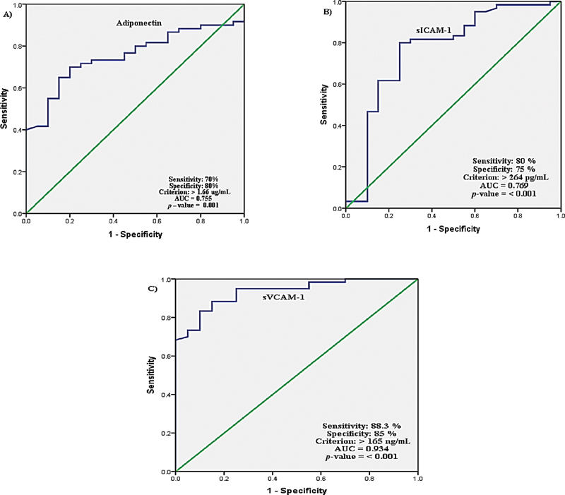
Fig 4: Receiver operating characteristic curve analyses for the diagnosis breast cancer from healthy controls. (A) Adiponectin. (B) soluble intercellular adhesion molecule-1 (sICAM-1). (C) Soluble vascular cell adhesion molecule-1 (sVCAM-1). AUC, area under the receiver operating characteristic (ROC) curve.
Discussion
There are common biomolecules which are increased and therefore are indicators for the future development of CV diseases and cancer; may be defined as oncovascular risk factors. Several biomolecules like cell adhesion molecules and adipocytokines may belong to this category as they are involved in the pathogenesis of cancer as well as CV diseases. Despite significant advancements in cancer research, BC remains a major public health concern and a top scientific research goal. BC is the most prevalent disease among women throughout nations and its fatality rates and incidence are expected to skyrocket in the future years.[13] Studies have shown that BC patients have higher levels of serum cytoadipokines, cell adhesion molecules, and increased levels of stress, anxiety, and depression.[2] [4] We assessed the changes in the levels of CV risk factors such as leptin, adiponectin, sICAM-1, sVCAM-1, and baFMD as well as the psychological distress in the progression of the disease to higher stages among the new patients diagnosed with BC.
The dyslipidemia observed in the present study could reflect the increasing requirement of cancer cells for cholesterol with the progression of the disease. Increased number of LDL receptors and increased activity of HMG-CoA reductase have been reported in cancerous cells which explains the increasing serum cholesterol levels.[14] Both glucose and lipid metabolism undergo changes to suit cancer progression.[15] [16] Higher quantities of lipids are needed for cancer proliferation for biosynthesis of signal molecules, biofilms, and organelles.[17] [18] Activation of cholesterol biosynthesis and derangement of lipid metabolism can be promoted by the tumor microenvironment.[19] The enhanced requirement of membrane components and signaling molecules like sphingomyelin and phosphatidyiinositol are met by increased lipid biosynthesis.[20] Administration of statins may prevent the progression of the disease. Although we avoided known diabetic patients from this study, there was an increase in fasting glucose levels within the normal range during the progression to higher stages of the disease. Increase in blood glucose levels promotes the proliferation of cancer cells. Several studies have already been initiated with metformin supplementation in the management of BC.[21]
Several epidemiologic studies have demonstrated a significant inverse association of serum adiponectin with BC risk.[22] However, few reports state that there was a significant association between high adiponectin level and increased risk for BC.[23] As per the report by Ye et al, adiponectin levels were not associated with the risk of BC in premenopausal women.[24] We found marginal increase in the levels of adiponectin with the increasing stages in our study. The increased adiponectin levels may protect against cancer progression. Adiponectin is secreted exclusively by adipose tissue.[5] It has antiatherogenic, insulin-sensitizing, and anti-inflammatory properties.[25] In women with a family history of BC, it has been reported that increased circulating levels of high molecular weight adiponectin is a risk factor.[26] It has been reported that adiponectin via fatty acid metabolic reprogramming triggers BC cell death.[27] Another study indicated that high adiponectin levels and insulin resistance may be associated with aging or low nutrition status.[28] This adiponectin paradox has yet to be clarified, which has hindered our understanding of the biological role of adiponectin. This way the increased expression of adiponectin observed in the study may be considered as a protective mechanism adopted by the body.
Leptin has been identified as a risk factor for BC among women.[29] In the present study, although we observed a trend for increase in the serum leptin levels, it did not reach statistical significance.
The levels of both sVCAM-1 and sICAM-1 were found to be increasing with the stages of the disease. Thielemann et al reported a similar association between the levels of these molecules and the increasing stages of the BC.[30] They found a correlation between the aggressiveness of the disease and the increasing concentrations of soluble cell adhesion molecules. In the present study, the ROC curves of adiponectin, sICAM-1, as well as sVCAM-1 were found to be useful in the diagnosis of patients with BC. The endothelial surface CAMs play an important role in the disease progression.[31] These proteins participate in the intercellular adhesion and in the adherence of leucocytes to activated vascular endothelial cells. These events are crucial in the extravasation and inflammation.[32] Inhibition of CAMs may arrest the progression of the disease. CAMs promote metastases to distant organs by facilitating the attachment of cancer cells to endothelial cells.[33] Soluble forms (sCAMs) are also present in the circulation.[32] The sCAMs are smaller than the cell surface parent molecules representing only their extracellular domains. The concentration of sCAMs in circulation is related to the activation of endothelial cells.[33] Also, we found significant association among adiponectin and cell adhesion molecules. This indicates the nexus among the oncovascular risk factors. The CV diseases and BC have common risk factors.[34] [35] The cell adhesion molecules are considered as CV risk factors.[36] Among the women who survive BC there is an increased probability for the development of CV diseases. Such individuals need to be recognized early enough to prevent future complications.
Not much information is available on the status of vascular endothelial function among the newly diagnosed patients with BC. In our study, we did not find any compromise in the endothelial function among the patients in comparison to healthy control. This information agrees with the previous report by Lee et al.[12]
As per the previous reports, BC patients have a significant level of psychological discomfort and are more prone to develop anxiety and depression.[37] We found significant increase in the levels of perceived stress, depression, and anxiety as revealed by the PSS, PHQ-9, and GAD-7 questionnaire scores with increasing stages of the disease progression among the patients with BC in comparison to healthy controls. Cancer can cause psychiatric issues, particularly in women with BC, and these effects can affect both the patients and their family members.[4] Fear of losing the breast, as well as real breast loss, can have a severe impact on women's sentiments of sexuality, parenting, body image, and attractiveness. Mastectomy is thought to strip a woman of her femininity, fertility, beauty, and desire, hurting her body image and, as a result, producing psychological issues in the patient. The two most frequent psychological comorbidities among BC patients are depression and anxiety. Following a BC diagnosis, anxiety is frequently comorbid with depression, and it may be exacerbated during chemotherapy.[37] Moderate to high associations were found in the present study between disease severity and stress scores, PSS, PHQ-9, and GAD-7. The level of stress, anxiety, and depression could potentially act as causes for the increased secretion of proinflammatory cytokines in advanced stages of BC.[38] We found weak to moderate association of adipocytokines and cell adhesion molecules with the scores of stress, anxiety, and depression. The observation that the PSS had the maximum independent association with disease progression among the Indian patients with cancer necessitates investigating the causes of stress among them.
Conclusion
Increment in the levels of oncovascular risk factors with the progressing stages of BC suggests that these patients may be at higher risk of developing CV disease in future. This may give us a window to identify candidates for early intervention which may help to reduce the incidence of CV diseases in future. Adiponectin, sICAM-1, and sVCAM-1 are useful in the diagnosis of BC. There was significant increase in the levels of perceived stress, depression, and anxiety scores with the increasing stages of disease progression among the patients with BC. There was moderate association among oncovascular risk factors, psychological distress levels, and disease progression of the patients with BC. This warrants appropriate psychological counseling for patients with BC for bringing forth a better treatment outcome.
Conflict of Interest
None declared.
Acknowledgment
We gratefully acknowledge the technical assistance rendered by Ms. Durga Devi in carrying out this study.
Patient Consent
Informed consent was obtained from the participants.
Authors' Contributions
G.A.: Experimental studies, data acquisition, data analysis, statistical analysis, and manuscript preparation.
Z.B.: Concept, design, definition of intellectual content, literature search, data acquisition, data analysis, statistical analysis, manuscript preparation, manuscript editing, and manuscript review.
P.G.: Concept, design, data acquisition, data acquisition, and manuscript review.
S.V.C.: Concept, design, data acquisition, and manuscript review.
K.T.: Manuscript preparation, manuscript editing, and manuscript review.
The manuscript has been read and approved by all the authors.
References
- Shah R, Rosso K, Nathanson SD. Pathogenesis, prevention, diagnosis and treatment of breast cancer. World J Clin Oncol 2014; 5 (03) 283-298
- Christodoulatos GS, Spyrou N, Kadillari J, Psallida S, Dalamaga M. The role of adipokines in breast cancer: current evidence and perspectives. Curr Obes Rep 2019; 8 (04) 413-433
- Kim BK, Lee JW, Park PJ. et al. The multiplex bead array approach to identifying serum biomarkers associated with breast cancer. Breast Cancer Res 2009; 11 (02) R22
- Alagizy HA, Soltan MR, Soliman SS, Hegazy NN, Gohar SF. Anxiety, depression and perceived stress among breast cancer patients: single institute experience. Middle East Curr Psychiat 2020; 27 (01) 29
- Assiri AMA, Kamel HFM, Hassanien MFR. Resistin, visfatin, adiponectin, and leptin: risk of breast cancer in pre- and postmenopausal Saudi females and their possible diagnostic and predictive implications as novel biomarkers. Dis Markers 2015; 2015: 253519
- Sánchez-Jiménez F, Pérez-Pérez A, de la Cruz-Merino L, Sánchez-Margalet V. Obesity and breast cancer: role of leptin. Front Oncol 2019; 9: 596
- Thielemann A, Baszczuk A, Kopczyński Z, Nowak A, Grodecka-Gazdecka S. The clinical usefulness of assessing the concentration of cell adhesion molecules sVCAM-1 and sICAM-1 in the serum of women with primary breast cancer. Contemp Oncol (Pozn) 2014; 18 (04) 252-259
- Pilevarzadeh M, Amirshahi M, Afsargharehbagh R, Rafiemanesh H, Hashemi S-M, Balouchi A. Global prevalence of depression among breast cancer patients: a systematic review and meta-analysis. Breast Cancer Res Treat 2019; 176 (03) 519-533
- Perceived stress scale.pdf [Internet]. Accessed August 11, 2019 at: https://das.nh.gov/wellness/docs/percieved stress scale.pdf
- Phqscreeners [Internet]. phqscreeners. Accessed October 10, 2019 at: https://www.phqscreeners.com/
- Spitzer RL, Kroenke K, Williams JBW, Löwe B. A brief measure for assessing generalized anxiety disorder: the GAD-7. Arch Intern Med 2006; 166 (10) 1092-1097
- Lee K, Kang I, Mack WJ. et al. Effects of high-intensity interval training on vascular endothelial function and vascular wall thickness in breast cancer patients receiving anthracycline-based chemotherapy: a randomized pilot study. Breast Cancer Res Treat 2019; 177 (02) 477-485
- Anastasiadi Z, Lianos GD, Ignatiadou E, Harissis HV, Mitsis M. Breast cancer in young women: an overview. Updates Surg 2017; 69 (03) 313-317
- Chimento A, Casaburi I, Avena P. et al. Cholesterol, and its metabolites in tumor growth: therapeutic potential of statins in cancer treatment. Front Endocrinol (Lausanne) 2019; 9: 807
- Corbet C, Feron O. Emerging roles of lipid metabolism in cancer progression. Curr Opin Clin Nutr Metab Care 2017; 20 (04) 254-260
- Corbet C, Pinto A, Martherus R, Santiago de Jesus JP, Polet F, Feron O. Acidosis drives the reprogramming of fatty acid metabolism in cancer cells through changes in mitochondrial and histone acetylation. Cell Metab 2016; 24 (02) 311-323
- Furuta E, Pai SK, Zhan R. et al. Fatty acid synthase gene is up-regulated by hypoxia via activation of Akt and sterol regulatory element binding protein-1. Cancer Res 2008; 68 (04) 1003-1011
- Lu S, Archer MC. Sp1 coordinately regulates de novo lipogenesis and proliferation in cancer cells. Int J Cancer 2010; 126 (02) 416-425
- Hanahan D, Weinberg RA. Hallmarks of cancer: the next generation. Cell 2011; 144 (05) 646-674
- Zeng Y, Ren K, Zhu X, Zheng Z, Yi G. Long noncoding RNAs: advances in lipid metabolism. Adv Clin Chem 2018; 87: 1-36
- Cejuela M, Martin-Castillo B, Menendez JA, Pernas S. Metformin and breast cancer: where are we now?. Int J Mol Sci 2022; 23 (05) 2705
- Yu Z, Tang S, Ma H, Duan H, Zeng Y. Association of serum adiponectin with breast cancer: a meta-analysis of 27 case-control studies. Medicine (Baltimore) 2019; 98 (06) e14359
- Liu LY, Wang M, Ma ZB. et al. The role of adiponectin in breast cancer: a meta-analysis. PLoS One 2013; 8 (08) e73183
- Ye J, Jia J, Dong S. et al. Circulating adiponectin levels and the risk of breast cancer: a meta-analysis. Eur J Cancer Prev 2014; 23 (03) 158-165
- Housa D, Housová J, Vernerová Z, Haluzík M. Adipocytokines and cancer. Physiol Res 2006; 55 (03) 233-244
- Guo MM, Duan XN, Cui SD. et al. Circulating high-molecular-weight (HMW) adiponectin level is related with breast cancer risk better than total adiponectin: a case-control study. PLoS One 2015; 10 (06) e0129246
- Pham DV, Park PH. Adiponectin triggers breast cancer cell death via fatty acid metabolic reprogramming. J Exp Clin Cancer Res 2022; 41 (01) 9
- Muratsu J, Kamide K, Fujimoto T. et al. The combination of high levels of adiponectin and insulin resistance are affected by aging in non-obese old peoples. Front Endocrinol (Lausanne) 2022; 12: 805244
- Pan H, Deng L-L, Cui J-Q. et al. Association between serum leptin levels and breast cancer risk: an updated systematic review and meta-analysis. Medicine (Baltimore) 2018; 97 (27) e11345
- Thielemann A, Baszczuk A, Kopczyński Z, Nowak A, Grodecka-Gazdecka S. The clinical usefulness of assessing the concentration of cell adhesion molecules sVCAM-1 and sICAM-1 in the serum of women with primary breast cancer. Contemp Oncol (Pozn) 2014; 18 (04) 252-259
-
Ilyas M. Adhesion molecule expression in breast cancer: the phoenix in tumour metastasis?. J Pathol 2000; 190 (01) 3-5 3.0.CO;2-5" target="linkout" data-target="CrossRef" data-id="CrossRef" data-url="https://doi.org/10.1002/(SICI)1096-9896(200001)190:1<3>3.0.CO;2-5" style="color: rgb(1, 52, 118); display: inline-block; padding: 5px 10px 5px 12px; line-height: 16.8px;">Crossref PubMed Search in Google ScholarDownload RIS citation
- Gho YS, Kim PN, Li HC, Elkin M, Kleinman HK. Stimulation of tumor growth by human soluble intercellular adhesion molecule-1. Cancer Res 2001; 61 (10) 4253-4257
- Pietruczuk M, Pietruczuk A, Pancewicz S, Hermanowska-Szpakowicz T. ICAM-1: structure, biological role and clinical significance [in Polish]. Pol Merkuriusz Lek 2004; 17 (101) 507-511
- Gulati M, Mulvagh SL. The connection between the breast and heart in a woman: Breast cancer and cardiovascular disease. Clin Cardiol 2018; 41 (02) 253-257
- Mehta LS, Watson KE, Barac A. et al; American Heart Association Cardiovascular Disease in Women and Special Populations Committee of the Council on Clinical Cardiology; Council on Cardiovascular and Stroke Nursing; and Council on Quality of Care and Outcomes Research. Cardiovascular disease and breast cancer: where these entities intersect: a scientific statement from the American Heart Association. Circulation 2018; 137 (08) e30-e66
- Peres BU, Hirsch Allen AJ, Daniele P. et al. Circulating levels of cell adhesion molecules and risk of cardiovascular events in obstructive sleep apnea. PLoS One 2021; 16 (07) e0255306
- Reece JC, Chan YF, Herbert J, Gralow J, Fann JR. Course of depression, mental health service utilization and treatment preferences in women receiving chemotherapy for breast cancer. Gen Hosp Psychiatry 2013; 35 (04) 376-381
- Li M, Kouzmina E, McCusker M. et al. Cytokines and depression in cancer patients and caregivers. Neuropsychiatr Dis Treat 2017; 13: 2903-2911
Address for correspondence
Publication History
Article published online:
17 September 2024
© 2024. The Author(s). This is an open access article published by Thieme under the terms of the Creative Commons Attribution License, permitting unrestricted use, distribution, and reproduction so long as the original work is properly cited. (https://creativecommons.org/licenses/by/4.0/)
Thieme Medical and Scientific Publishers Private Limited
A-13A, Graphix Tower 1, 6th floor, Sector 62, Noida 201309, Uttar Pradesh, India
Fig 1:Comparison of soluble vascular cell adhesion molecule-1 (sVCAM-1) values between the study groups. (A) Control vs. stage 1 and 2; p < 0.001. (B) Control vs. stage 3; p < 0.001. (C) Control vs. stage 4; p < 0.001.
Fig 2: Comparison of Perceived Stress Scale (PSS) scores among the different stages of breast cancer in comparison to healthy controls. (A) Control vs. stage 1 and 2; p < 0.001. (B) Control vs. stage 3; p < 0.001. (C) Control vs. stage 4; p < 0.001.
Fig 3: Comparison of Patient Health Questionnaire-9 (PHQ-9) scores among the different stages of breast cancer in comparison to healthy controls. (A) Control vs. stage 1 and 2; p = 0 0.003. (B) Control vs. stage 3; p < 0.001. (C) Control vs. stage 4; p < 0.001.
Fig 4: Comparison of Generalized Anxiety Disorder-7 (GAD-7) scores among the different stages of breast cancer in comparison to healthy controls. (A) Control vs. stage 1 and 2; p = 0.002. (B) Control vs. stage 3; p = 0.002. (C) Control vs. stage 4; p < 0.001. (D) Stage 1 and 2 vs. stage 4; p = 0.041. (E) Stage 3 vs. stage 4; p = 0.042.
Fig 4: Receiver operating characteristic curve analyses for the diagnosis breast cancer from healthy controls. (A) Adiponectin. (B) soluble intercellular adhesion molecule-1 (sICAM-1). (C) Soluble vascular cell adhesion molecule-1 (sVCAM-1). AUC, area under the receiver operating characteristic (ROC) curve.
References
- Shah R, Rosso K, Nathanson SD. Pathogenesis, prevention, diagnosis and treatment of breast cancer. World J Clin Oncol 2014; 5 (03) 283-298
- Christodoulatos GS, Spyrou N, Kadillari J, Psallida S, Dalamaga M. The role of adipokines in breast cancer: current evidence and perspectives. Curr Obes Rep 2019; 8 (04) 413-433
- Kim BK, Lee JW, Park PJ. et al. The multiplex bead array approach to identifying serum biomarkers associated with breast cancer. Breast Cancer Res 2009; 11 (02) R22
- Alagizy HA, Soltan MR, Soliman SS, Hegazy NN, Gohar SF. Anxiety, depression and perceived stress among breast cancer patients: single institute experience. Middle East Curr Psychiat 2020; 27 (01) 29
- Assiri AMA, Kamel HFM, Hassanien MFR. Resistin, visfatin, adiponectin, and leptin: risk of breast cancer in pre- and postmenopausal Saudi females and their possible diagnostic and predictive implications as novel biomarkers. Dis Markers 2015; 2015: 253519
- Sánchez-Jiménez F, Pérez-Pérez A, de la Cruz-Merino L, Sánchez-Margalet V. Obesity and breast cancer: role of leptin. Front Oncol 2019; 9: 596
- Thielemann A, Baszczuk A, Kopczyński Z, Nowak A, Grodecka-Gazdecka S. The clinical usefulness of assessing the concentration of cell adhesion molecules sVCAM-1 and sICAM-1 in the serum of women with primary breast cancer. Contemp Oncol (Pozn) 2014; 18 (04) 252-259
- Pilevarzadeh M, Amirshahi M, Afsargharehbagh R, Rafiemanesh H, Hashemi S-M, Balouchi A. Global prevalence of depression among breast cancer patients: a systematic review and meta-analysis. Breast Cancer Res Treat 2019; 176 (03) 519-533
- Perceived stress scale.pdf [Internet]. Accessed August 11, 2019 at: https://das.nh.gov/wellness/docs/percieved stress scale.pdf
- Phqscreeners [Internet]. phqscreeners. Accessed October 10, 2019 at: https://www.phqscreeners.com/
- Spitzer RL, Kroenke K, Williams JBW, Löwe B. A brief measure for assessing generalized anxiety disorder: the GAD-7. Arch Intern Med 2006; 166 (10) 1092-1097
- Lee K, Kang I, Mack WJ. et al. Effects of high-intensity interval training on vascular endothelial function and vascular wall thickness in breast cancer patients receiving anthracycline-based chemotherapy: a randomized pilot study. Breast Cancer Res Treat 2019; 177 (02) 477-485
- Anastasiadi Z, Lianos GD, Ignatiadou E, Harissis HV, Mitsis M. Breast cancer in young women: an overview. Updates Surg 2017; 69 (03) 313-317
- Chimento A, Casaburi I, Avena P. et al. Cholesterol, and its metabolites in tumor growth: therapeutic potential of statins in cancer treatment. Front Endocrinol (Lausanne) 2019; 9: 807
- Corbet C, Feron O. Emerging roles of lipid metabolism in cancer progression. Curr Opin Clin Nutr Metab Care 2017; 20 (04) 254-260
- Corbet C, Pinto A, Martherus R, Santiago de Jesus JP, Polet F, Feron O. Acidosis drives the reprogramming of fatty acid metabolism in cancer cells through changes in mitochondrial and histone acetylation. Cell Metab 2016; 24 (02) 311-323
- Furuta E, Pai SK, Zhan R. et al. Fatty acid synthase gene is up-regulated by hypoxia via activation of Akt and sterol regulatory element binding protein-1. Cancer Res 2008; 68 (04) 1003-1011
- Lu S, Archer MC. Sp1 coordinately regulates de novo lipogenesis and proliferation in cancer cells. Int J Cancer 2010; 126 (02) 416-425
- Hanahan D, Weinberg RA. Hallmarks of cancer: the next generation. Cell 2011; 144 (05) 646-674
- Zeng Y, Ren K, Zhu X, Zheng Z, Yi G. Long noncoding RNAs: advances in lipid metabolism. Adv Clin Chem 2018; 87: 1-36
- Cejuela M, Martin-Castillo B, Menendez JA, Pernas S. Metformin and breast cancer: where are we now?. Int J Mol Sci 2022; 23 (05) 2705
- Yu Z, Tang S, Ma H, Duan H, Zeng Y. Association of serum adiponectin with breast cancer: a meta-analysis of 27 case-control studies. Medicine (Baltimore) 2019; 98 (06) e14359
- Liu LY, Wang M, Ma ZB. et al. The role of adiponectin in breast cancer: a meta-analysis. PLoS One 2013; 8 (08) e73183
- Ye J, Jia J, Dong S. et al. Circulating adiponectin levels and the risk of breast cancer: a meta-analysis. Eur J Cancer Prev 2014; 23 (03) 158-165
- Housa D, Housová J, Vernerová Z, Haluzík M. Adipocytokines and cancer. Physiol Res 2006; 55 (03) 233-244
- Guo MM, Duan XN, Cui SD. et al. Circulating high-molecular-weight (HMW) adiponectin level is related with breast cancer risk better than total adiponectin: a case-control study. PLoS One 2015; 10 (06) e0129246
- Pham DV, Park PH. Adiponectin triggers breast cancer cell death via fatty acid metabolic reprogramming. J Exp Clin Cancer Res 2022; 41 (01) 9
- Muratsu J, Kamide K, Fujimoto T. et al. The combination of high levels of adiponectin and insulin resistance are affected by aging in non-obese old peoples. Front Endocrinol (Lausanne) 2022; 12: 805244
- Pan H, Deng L-L, Cui J-Q. et al. Association between serum leptin levels and breast cancer risk: an updated systematic review and meta-analysis. Medicine (Baltimore) 2018; 97 (27) e11345
- Thielemann A, Baszczuk A, Kopczyński Z, Nowak A, Grodecka-Gazdecka S. The clinical usefulness of assessing the concentration of cell adhesion molecules sVCAM-1 and sICAM-1 in the serum of women with primary breast cancer. Contemp Oncol (Pozn) 2014; 18 (04) 252-259
-
Ilyas M. Adhesion molecule expression in breast cancer: the phoenix in tumour metastasis?. J Pathol 2000; 190 (01) 3-5 3.0.CO;2-5" target="linkout" data-target="CrossRef" data-id="CrossRef" data-url="https://doi.org/10.1002/(SICI)1096-9896(200001)190:1<3>3.0.CO;2-5" style="color: rgb(1, 52, 118); display: inline-block; padding: 5px 10px 5px 12px; line-height: 16.8px;">Crossref PubMed Search in Google ScholarDownload RIS citation
- Gho YS, Kim PN, Li HC, Elkin M, Kleinman HK. Stimulation of tumor growth by human soluble intercellular adhesion molecule-1. Cancer Res 2001; 61 (10) 4253-4257
- Pietruczuk M, Pietruczuk A, Pancewicz S, Hermanowska-Szpakowicz T. ICAM-1: structure, biological role and clinical significance [in Polish]. Pol Merkuriusz Lek 2004; 17 (101) 507-511
- Gulati M, Mulvagh SL. The connection between the breast and heart in a woman: Breast cancer and cardiovascular disease. Clin Cardiol 2018; 41 (02) 253-257
- Mehta LS, Watson KE, Barac A. et al; American Heart Association Cardiovascular Disease in Women and Special Populations Committee of the Council on Clinical Cardiology; Council on Cardiovascular and Stroke Nursing; and Council on Quality of Care and Outcomes Research. Cardiovascular disease and breast cancer: where these entities intersect: a scientific statement from the American Heart Association. Circulation 2018; 137 (08) e30-e66
- Peres BU, Hirsch Allen AJ, Daniele P. et al. Circulating levels of cell adhesion molecules and risk of cardiovascular events in obstructive sleep apnea. PLoS One 2021; 16 (07) e0255306
- Reece JC, Chan YF, Herbert J, Gralow J, Fann JR. Course of depression, mental health service utilization and treatment preferences in women receiving chemotherapy for breast cancer. Gen Hosp Psychiatry 2013; 35 (04) 376-381
- Li M, Kouzmina E, McCusker M. et al. Cytokines and depression in cancer patients and caregivers. Neuropsychiatr Dis Treat 2017; 13: 2903-2911