 PDF
PDF  Views
Views  Share
Share


Isolated conjunctival mass presenting as acute myeloid leukemia in an infant
CC BY-NC-ND 4.0 · Indian J Med Paediatr Oncol 2016; 37(02): 124
DOI: DOI: 10.4103/0971-5851.180146
Publication History
Article published online:
12 July 2021
© 2016. Indian Society of Medical and Paediatric Oncology. This is an open access article published by Thieme under the terms of the Creative Commons Attribution-NonDerivative-NonCommercial-License, permitting copying and reproduction so long as the original work is given appropriate credit. Contents may not be used for commercial purposes, or adapted, remixed, transformed or built upon. (https://creativecommons.org/licenses/by-nc-nd/4.0/.)
Thieme Medical and Scientific Publishers Pvt. Ltd.
A-12, 2nd Floor, Sector 2, Noida-201301 UP, India
Sir,
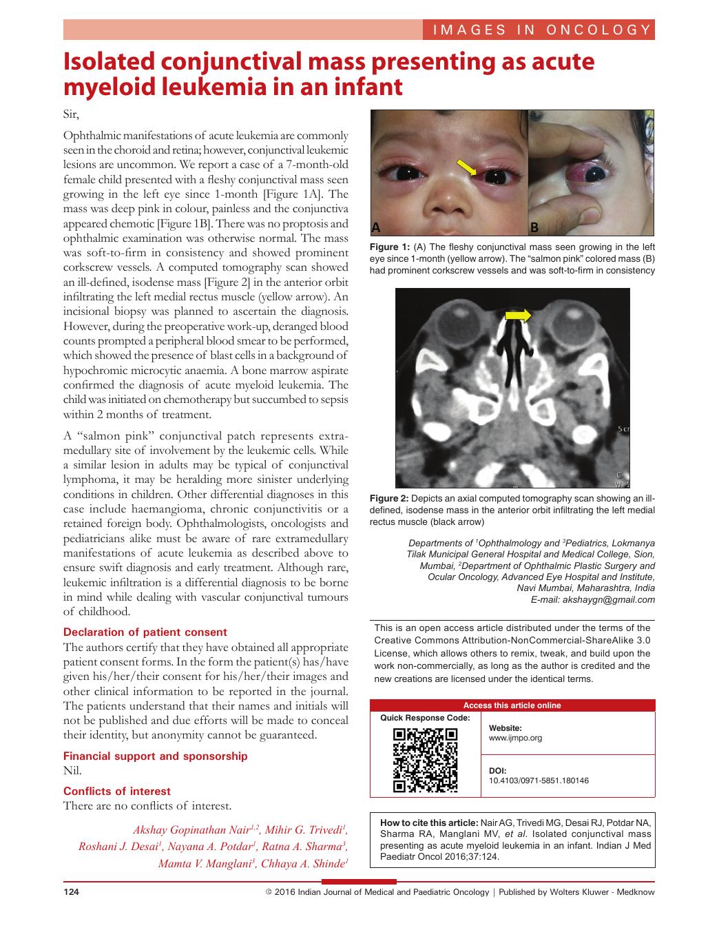
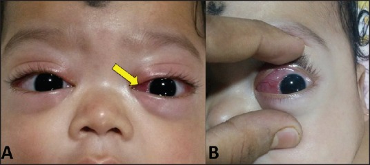
Ophthalmic manifestations of acute leukemia are commonly seen in the choroid and retina; however, conjunctival leukemic lesions are uncommon. We report a case of a 7-month-old female child presented with a fleshy conjunctival mass seen growing in the left eye since 1-month [Figure 1A]. The mass was deep pink in colour, painless and the conjunctiva appeared chemotic [Figure 1B]. There was no proptosis and ophthalmic examination was otherwise normal. The mass was soft-to-firm in consistency and showed prominent corkscrew vessels. A computed tomography scan showed an ill-defined, isodense mass [Figure 2] in the anterior orbit infiltrating the left medial rectus muscle (yellow arrow). An incisional biopsy was planned to ascertain the diagnosis. However, during the preoperative work-up, deranged blood counts prompted a peripheral blood smear to be performed, which showed the presence of blast cells in a background of hypochromic microcytic anaemia. A bone marrow aspirate confirmed the diagnosis of acute myeloid leukemia. The child was initiated on chemotherapy but succumbed to sepsis within 2 months of treatment.
| Fig. 1 (A) The fleshy conjunctival mass seen growing in the left eye since 1-month (yellow arrow). The “salmon pink” colored mass (B) had prominent corkscrew vessels and was soft-to-firm in consistency
| Fig. 2 Depicts an axial computed tomography scan showing an ill-defined, isodense mass in the anterior orbit infiltrating the left medial rectus muscle (black arrow)
A “salmon pink” conjunctival patch represents extra-medullary site of involvement by the leukemic cells. While a similar lesion in adults may be typical of conjunctival lymphoma, it may be heralding more sinister underlying conditions in children. Other differential diagnoses in this case include haemangioma, chronic conjunctivitis or a retained foreign body. Ophthalmologists, oncologists and pediatricians alike must be aware of rare extramedullary manifestations of acute leukemia as described above to ensure swift diagnosis and early treatment. Although rare, leukemic infiltration is a differential diagnosis to be borne in mind while dealing with vascular conjunctival tumours of childhood.
Declaration of patient consent
The authors certify that they have obtained all appropriate patient consent forms. In the form the patient(s) has/have given his/her/their consent for his/her/their images and other clinical information to be reported in the journal. The patients understand that their names and initials will not be published and due efforts will be made to conceal their identity, but anonymity cannot be guaranteed.
Financial support and sponsorship
Nil.
Conflicts of interest
There are no conflicts of interest.
| Fig. 1 (A) The fleshy conjunctival mass seen growing in the left eye since 1-month (yellow arrow). The “salmon pink” colored mass (B) had prominent corkscrew vessels and was soft-to-firm in consistency