 PDF
PDF  Views
Views  Share
Share


Late Recurrence of Malignant Choroidal Melanoma Mimicking Hepatocellular Carcinoma
CC BY-NC-ND 4.0 ? Indian J Med Paediatr Oncol 2018; 39(01): 109-110
DOI: DOI: 10.4103/ijmpo.ijmpo_12_16
Abstract
Late recurrence (after more than 10 years) of choroidal melanoma, though infrequent, has been documented well in Western countries. In India, however, this disease being uncommon, there is no data regarding its late recurrence. Here, we report a case of late metastatic manifestation of choroidal melanoma in the form of liver metastases in a 45-year-old man who presented with abdominal pain; metastases to liver occurred 10 years after enucleation of the right eye.
Keywords
Choroidal melanoma - India - late recurrence
Publication History
23 June 2021
A-12, 2nd Floor, Sector 2, Noida-201301 UP, India
Abstract
Late recurrence (after more than 10 years) of choroidal melanoma, though infrequent, has been documented well in Western countries. In India, however, this disease being uncommon, there is no data regarding its late recurrence. Here, we report a case of late metastatic manifestation of choroidal melanoma in the form of liver metastases in a 45-year-old man who presented with abdominal pain; metastases to liver occurred 10 years after enucleation of the right eye.
Keywords
Choroidal melanoma - India - late recurrence
Introduction
Choroidal Melanoma is the most common primary intraocular malignant tumor in western countries but is rare in India. Malignant melanomas are known for their late recurrences. Most common site of recurrence is liver. But being a rare tumor, late recurrence with metastasis to liver has not been reported from India.
Case Report
A 45-year-old man presented to us with complaints of pain in the abdomen with heaviness at right hypochondrium. Examination revealed hepatomegaly extending up to 5 cm below the right costal margin. On palpation, the liver was hard and nontender.
Ultrasonography (USG) and computed tomography scan revealed enlarged liver which measured approximately 25 cm in craniocaudal span. Multiple arterial phase enhancing heterogeneous nodular lesions showing venous washout were scattered in both lobes. Largest of these measures approximately 13 cm ? 11 cm ? 10 cm involving segment VIII and shows internal calcific foci. Subtle left lobar intrahepatic biliary radical prominence is noted with a simple 11 mm cyst in segment II. Furthermore, few enlarged heterogeneously enhancing periportal lymph nodes were seen. These appear to be extrinsically compressing the common hepatic duct [Figure 1]. In view of arterial enhancement, of hypervascular metastases from occult primary, neuroendocrine tumor (NET) with liver secondaries or multifocal hepatocellualr carcinoma (HCC) was the suggested differential diagnosis.
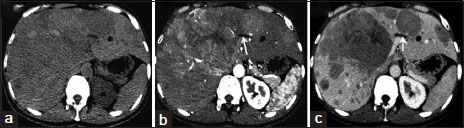
|?Figure.1Axial images of multiphase computed tomography scan of the upper abdomen revealed multiple hypodense lesions of variable size involving both lobe of the liver (a), Which shows hyperenhancement in hepatic arterial phase (b) and washout in subsequent portal venous phase (c). One of the large central mass is having central nonenhancing area suggestive of necrosis and also causing mild dilatation of the left hepatic duct (white arrows)
Serum tumor markers (AFP, CEA, and CA19.9) were normal. USG-guided biopsy of the lesion revealed tumor cells arranged in nodules separated by fibro-collagenous stroma. The tumor cells were round to spindle shaped and showed coarse nuclear chromatin, porminent eosinophilic nucleoli, and moderate amount of cytoplasm containing coarse granular black melanin pigment suggestive of melanoma [Figure 2].
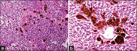
|?Figure.2(a) Malignant pleomorphic cells with brownish black intracytoplasmic melanin pigment H and E, ?200. (b) Tumor cells with brownish black melanin pigment H and E, ?400
A detailed history retrospectively revealed a history of choroidal melanoma for which enucleation was done 10 years prior.
Discussion
Choroidal melanoma is rare in dark pigmented races and has been reported infrequently in Indian population. Its incidence was reported <0 href="https://www.thieme-connect.com/products/ejournals/html/10.4103/ijmpo.ijmpo_12_16#JR_1" xss=removed>1] It causes death, almost always secondary to distant metastases. Principal target organ for metastasis is liver, with almost 50% of the patients developing liver metastases up to 15 years after diagnosis.[2]
The reason behind this target organ is likely due to increased expression of c-Met, IGF-IR, and CXCR4 in Uveal Melanoma cells as compared to normal melanocytes. The liver is the only organ that highly expresses the corresponding ligands of these receptors (HGF, IGF, and CXCL12), indicating that these pathways may be highly involved in the liver-specific metastases in Uveal melanoma.[2]
As melanoma is uncommon tumor; very few late recurrences have been reported from India.[3],[4],[5] In fact, it is a lost history about previous ophthalmic intervention due to its rarity; which is why, the differential diagnosis on radiology were kept as hypervascular metastasis from unknown primary, NET or HCC; more common entities than melanoma. Choroidal melanoma is rare and masquerading that it is easily misdiagnosed. On his first visit, the patient gave a history of ophthalmic surgery, 10 years back and he was not aware of the cause and did not have histopathological report at that time. We asked for the previous histopathology report after the present biopsy suggestive of melanoma. Here, the relevance of history comes which signifies its late recurrence and thereby its prognosis. This case also highlights the requirement of long-term follow-up in melanoma patients, as the incidence of late recurrence in melanoma is 6.8?cording to western literature.[6]
Conclusion
This is first case report from India, highlighting a choroidal melanoma presenting with liver metastases after 10 years of enucleation.
Conflict of Interest
There are no conflicts of interest.
References
- Boyle P, Maisonneuve P, Dor? JF.?Epidemiology of malignant melanoma. Br Med Bull 1995; 51: 523-47
- Bakalian S, Marshall JC, Logan P, Faingold D, Maloney S, Di CesareS.?et al.?Molecular pathways mediating liver metastasis in patients with uveal melanoma. Clin Cancer Res 2008; 14: 951-6
- Agarwal SR, Bhattacharya I, Patil YV, Amrapurkar AD.?Choroidal melanoma metastatizing to the biliary system: A diagnostic dilemma. Indian J Med Paediatr Oncol 2009; 30: 138-40
- Gowrinath K, Geetha V.?Late recurrence of malignant melanoma presenting with hemoptysis. Lung India 2010; 27: 178-80
- Singh P, Singh A.?Choroidal melanoma. Oman J Ophthalmol 2012; 5: 3-9
- Faries MB, Steen S, Ye X, Sim M, Morton DL.?Late recurrence in melanoma: Clinical implications of lost dormancy. J Am Coll Surg 2013; 217: 27-34
Address for correspondence
Publication History
Article published online:
23 June 2021
? 2018. Indian Society of Medical and Paediatric Oncology. This is an open access article published by Thieme under the terms of the Creative Commons Attribution-NonDerivative-NonCommercial-License, permitting copying and reproduction so long as the original work is given appropriate credit. Contents may not be used for commercial purposes, or adapted, remixed, transformed or built upon. (https://creativecommons.org/licenses/by-nc-nd/4.0/.)
Thieme Medical and Scientific Publishers Pvt. Ltd.
A-12, 2nd Floor, Sector
2, Noida-201301 UP, India
|?Figure.1Axial images of multiphase computed tomography scan of the upper abdomen revealed multiple hypodense lesions of variable size involving both lobe of the liver (a), Which shows hyperenhancement in hepatic arterial phase (b) and washout in subsequent portal venous phase (c). One of the large central mass is having central nonenhancing area suggestive of necrosis and also causing mild dilatation of the left hepatic duct (white arrows)
|?Figure.2(a) Malignant pleomorphic cells with brownish black intracytoplasmic melanin pigment H and E, ?200. (b) Tumor cells with brownish black melanin pigment H and E, ?400
References
- Boyle P, Maisonneuve P, Dor? JF.?Epidemiology of malignant melanoma. Br Med Bull 1995; 51: 523-47
- Bakalian S, Marshall JC, Logan P, Faingold D, Maloney S, Di CesareS.?et al.?Molecular pathways mediating liver metastasis in patients with uveal melanoma. Clin Cancer Res 2008; 14: 951-6
- Agarwal SR, Bhattacharya I, Patil YV, Amrapurkar AD.?Choroidal melanoma metastatizing to the biliary system: A diagnostic dilemma. Indian J Med Paediatr Oncol 2009; 30: 138-40
- Gowrinath K, Geetha V.?Late recurrence of malignant melanoma presenting with hemoptysis. Lung India 2010; 27: 178-80
- Singh P, Singh A.?Choroidal melanoma. Oman J Ophthalmol 2012; 5: 3-9
- Faries MB, Steen S, Ye X, Sim M, Morton DL.?Late recurrence in melanoma: Clinical implications of lost dormancy. J Am Coll Surg 2013; 217: 27-34