 PDF
PDF  Views
Views  Share
Share


Microfilariasis of the breast mimicking malignancy
CC BY-NC-ND 4.0 · Indian J Med Paediatr Oncol 2013; 34(01): 47
DOI: DOI: 10.4103/0971-5851.113433
Abstract
We report a 65-year-old female who presented with a 3.5x3 cm retro-areolar lump of the right breast with associated right axillary lymphadenopathy, mimicking breast cancer. Mammography showed a well-defined mass in the central quadrant of the right breast. Fine needle aspiration cytology from the breast lump demonstrated many microfilariae of Wuchereria bancrofti.
Publication History
Article published online:
20 July 2021
© 2013. Indian Society of Medical and Paediatric Oncology. This is an open access article published by Thieme under the terms of the Creative Commons Attribution-NonDerivative-NonCommercial-License, permitting copying and reproduction so long as the original work is given appropriate credit. Contents may not be used for commercial purposes, or adapted, remixed, transformed or built upon. (https://creativecommons.org/licenses/by-nc-nd/4.0/.)
Thieme Medical and Scientific Publishers Pvt. Ltd.
A-12, 2nd Floor, Sector 2, Noida-201301 UP, India
Abstract
We report a 65-year-old female who presented with a 3.5×3 cm retro-areolar lump of the right breast with associated right axillary lymphadenopathy, mimicking breast cancer. Mammography showed a well-defined mass in the central quadrant of the right breast. Fine needle aspiration cytology from the breast lump demonstrated many microfilariae of Wuchereria bancrofti.
A 65-year-old female from Bihar presented with a complaint of a painless lump in the right breast for the last one year. On examination, a 3.5 × 3 cm firm, mobile, retroareolar lump was found in the right breast, along with a 1 × 1 cm single, firm, mobile lymph node in the right axilla. No skin change, nipple retraction, or peau d’orange was seen. Mammography showed a well-defined mass in the central quadrant of the right breast [Figure 1]. A clinical suspicion of breast carcinoma was considered, but fine-needle aspiration cytology (FNAC) from the lump revealed numerous sheathed microfilariae of Wuchereria bancrofti with rounded anterior and tapering posterior ends with elongated terminal nuclei, in the background of inflammatory cells [Figure 2]. No malignant cells were identified. FNAC from the right axillary lymph node showed reactive lymphocytosis. A midnight peripheral blood sample failed to show any microfilaria, even after diethylcarbamazine citrate (DEC) provocation. A diagnosis of occult microfilariasis of the right breast was made.
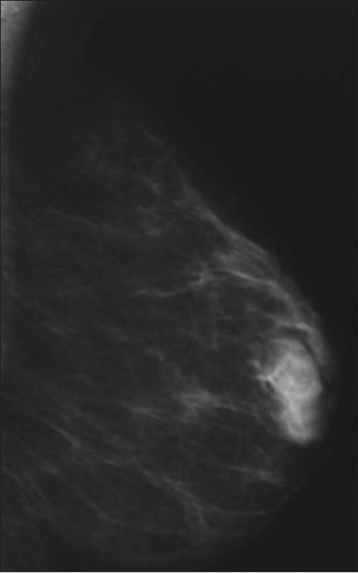
| Fig. 1 Mammography of the right breast (mediolateral oblique view) showing a well-defined mass in the central quadrant without any microcalcification
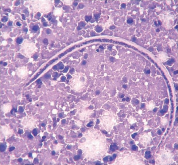
| Fig. 2 Photomicrograph showing a microfilaria of Wuchereria bancrofti with rounded anterior and tapered posterior ends, in the background of inflammatory cells (MGG ×40)
Footnotes
Source of Support: Nil
Conflict of Interest: None declared.
| Fig. 1 Mammography of the right breast (mediolateral oblique view) showing a well-defined mass in the central quadrant without any microcalcification
| Fig. 2 Photomicrograph showing a microfilaria of Wuchereria bancrofti with rounded anterior and tapered posterior ends, in the background of inflammatory cells (MGG ×40)