 PDF
PDF  Views
Views  Share
Share


Orbital and infratemporal fossa metastasis: An unusual initial presentation of adenocarcinoma of lung
CC BY-NC-ND 4.0 · Indian J Med Paediatr Oncol 2013; 34(02): 132-133
DOI: DOI: 10.4103/0971-5851.116221
Abstract
Orbital metastasis as initial presentation of adenocarcinoma of lung is an extremely rare phenomenon. Here, we report a 46-year-old non-smoker Asian woman, who presented with right eye proptosis due to right orbital and infratemporal fossa metastasis, as the first presentation of adenocarcinoma of right lung.
Publication History
Article published online:
20 July 2021
© 2013. Indian Society of Medical and Paediatric Oncology. This is an open access article published by Thieme under the terms of the Creative Commons Attribution-NonDerivative-NonCommercial-License, permitting copying and reproduction so long as the original work is given appropriate credit. Contents may not be used for commercial purposes, or adapted, remixed, transformed or built upon. (https://creativecommons.org/licenses/by-nc-nd/4.0/.)
Thieme Medical and Scientific Publishers Pvt. Ltd.
A-12, 2nd Floor, Sector 2, Noida-201301 UP, India
Abstract
Orbital metastasis as initial presentation of adenocarcinoma of lung is an extremely rare phenomenon. Here, we report a 46-year-old non-smoker Asian woman, who presented with right eye proptosis due to right orbital and infratemporal fossa metastasis, as the first presentation of adenocarcinoma of right lung.
CASE REPORT
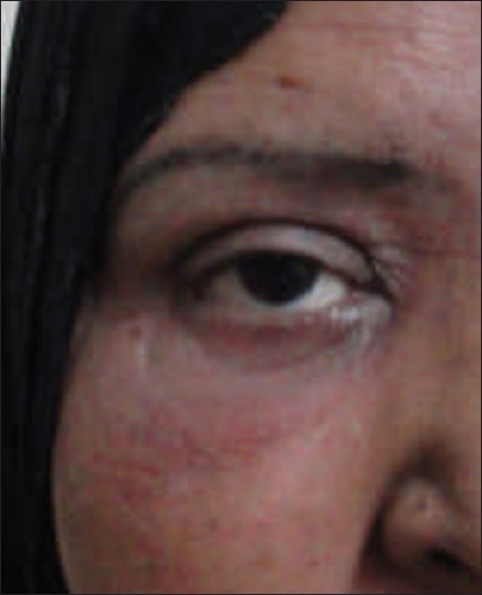
Orbital and infratemporal fossa (ITF) metastasis as initial presentation of adenocarcinoma of lung is extremely rare. We report a 46-year-old Asian woman, who presented with right eye proptosis and diffuse swelling over right cheek [Figure 1]. She had no other complaints or abnormal findings on clinical examination. A contrast enhanced computed tomographic (CECT) scan of face and neck revealed a solid mass in the right orbit with extension to the ITF [Figure 2]. Histopathologic examination of the biopsy from the mass showed poorly differentiated metastatic adenocarcinoma [Figure 3], which was thyroid transcription factor 1-positive [Figure 4]. A total-body CECT scan revealed a large heterogeneously enhancing mass lesion in the upper and middle lobe of the right lung [Figure 5]. CT guided biopsy from the lung mass revealed poorly differentiated adenocarcinoma [Figure 6]. No other metastases were detected. Therefore, she was diagnosed as a case of metastatic lung adenocarcinoma (T3N0M1, stage IV), and planned for palliative chemotherapy with pemetrexed (500 mg/m2) plus carboplatin (AUC = 5), iv on Day 1, every 3 weekly. She received 30 Gray/15 fraction palliative radiotherapy to the orbital mass. One month after completion of radiation, there was complete reduction of proptosis [Figure 7]. But, CECT thorax, done after 3 cycles of chemotherapy, revealed clear progression of the primary lung mass. The patient died 5 weeks later as a result of respiratory failure.
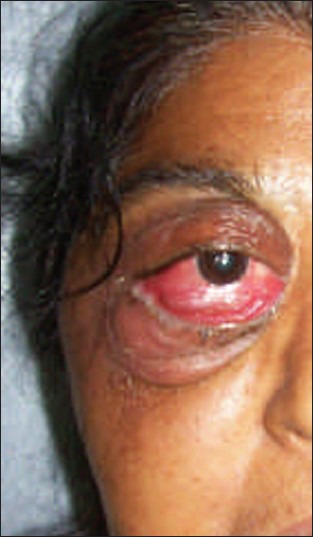
| Fig. 1 Initial presentation with right eye proptosis and right cheek swelling
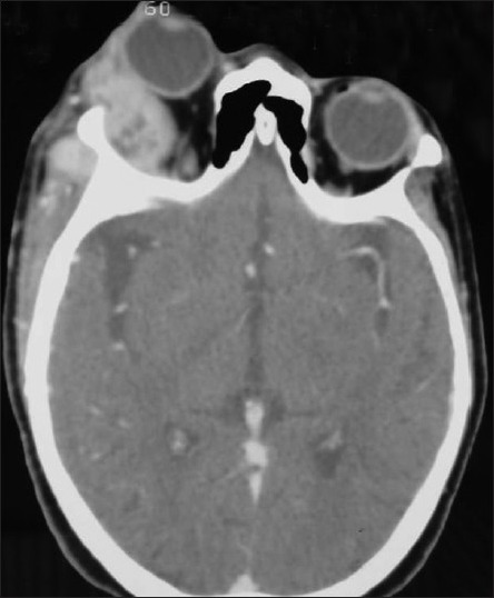
| Fig. 2 CECT scan of face and neck showing a solid mass in the right orbit with extension to the ITF
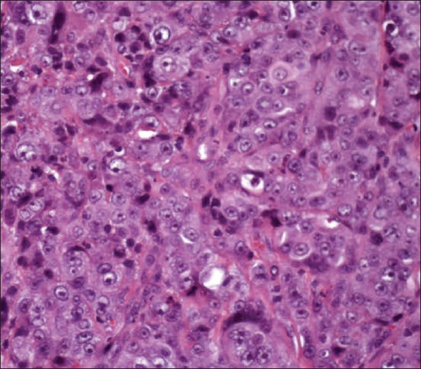
| Fig. 3 Histopathologic study of the orbital mass showing proliferation of adenocarcinoma cells (H and E, ×400)
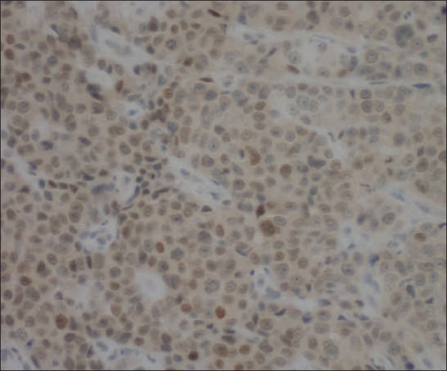
| Fig. 4 The tumor cells were positive for thyroid transcription factor 1 (×400)
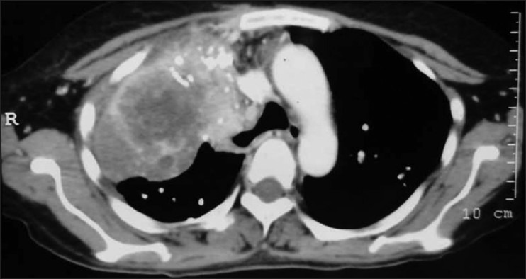
| Fig. 5 CECT scan of thorax showing large heterogeneously enhancing mass lesion in the upper and middle lobe of the right lung
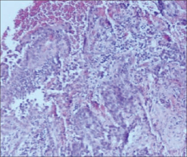
| Fig. 6 Histopathologic study of the CT guided biopsy from the lung mass, showing poorly differentiated adenocarcinoma (H and E, ×100)
| Fig. 7 One month post radiation, complete reduction of right eye proptosis
Footnotes
Source of Support: Nil
Conflict of Interest: None declared.
| Fig. 1 Initial presentation with right eye proptosis and right cheek swelling
| Fig. 2 CECT scan of face and neck showing a solid mass in the right orbit with extension to the ITF
| Fig. 3 Histopathologic study of the orbital mass showing proliferation of adenocarcinoma cells (H and E, ×400)
| Fig. 4 The tumor cells were positive for thyroid transcription factor 1 (×400)
| Fig. 5 CECT scan of thorax showing large heterogeneously enhancing mass lesion in the upper and middle lobe of the right lung
| Fig. 6 Histopathologic study of the CT guided biopsy from the lung mass, showing poorly differentiated adenocarcinoma (H and E, ×100)
| Fig. 7 One month post radiation, complete reduction of right eye proptosis