 PDF
PDF  Views
Views  Share
Share


Transformation of follicular lymphoma to high-grade Burkitt?s like lymphoma and acute lymphoblastic leukemia-L3 type
CC BY-NC-ND 4.0 · Indian J Med Paediatr Oncol 2013; 34(02): 136-137
DOI: DOI: 10.4103/0971-5851.116223
Publication History
Article published online:
20 July 2021
© 2013. Indian Society of Medical and Paediatric Oncology. This is an open access article published by Thieme under the terms of the Creative Commons Attribution-NonDerivative-NonCommercial-License, permitting copying and reproduction so long as the original work is given appropriate credit. Contents may not be used for commercial purposes, or adapted, remixed, transformed or built upon. (https://creativecommons.org/licenses/by-nc-nd/4.0/.)
Thieme Medical and Scientific Publishers Pvt. Ltd.
A-12, 2nd Floor, Sector 2, Noida-201301 UP, India
Sir,
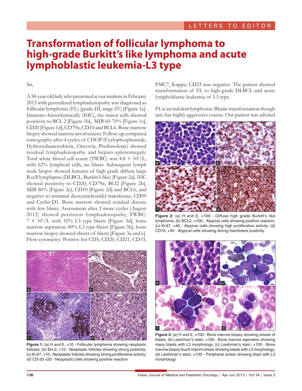
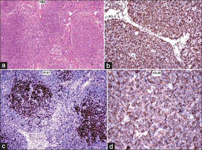
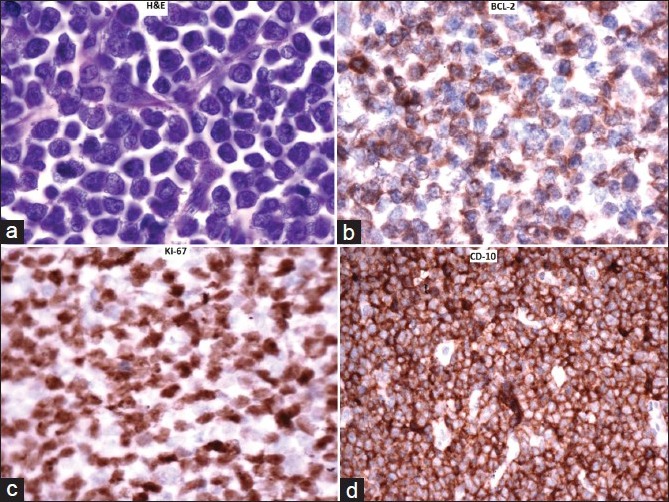
A 38-year-old lady who presented at our institute in February 2012 with generalized lymphadenopathy was diagnosed as follicular lymphoma (FL) (grade-III, stage-IV) [Figure 1a]. Immuno–histochemically (IHC), the tumor cells showed positivity to BCL 2 [Figure 1b], MIB 60-70% [Figure 1c], CD20 [Figure 1d], CD79α, CD10 and BCL6. Bone marrow biopsy showed marrow involvement. Follow-up computed tomography after 4 cycles of CHOP (Cyclophosphamide, Hydroxydaunorubicin, Oncovin, Prednisolone) showed residual lymphadenopathy and hepato-splenomegaly. Total white blood cell count (TWBC) was 4.8 × 10⁶/L, with 62% lymphoid cells, no blasts. Subsequent lymph node biopsy showed features of high grade diffuse large B cell lymphpma (DLBCL, Burkitt's like) [Figure 2a]. IHC showed positivity to CD20, CD79α, BCl2 [Figure 2b], MIB 80% [Figure 2c], CD10 [Figure 2d] and BCL6, and negative to terminal deoxynucleotidyl transferase, CD99 and Cyclin-D1. Bone marrow showed residual disease with few blasts. Assessment after 2 more cycles (August 2012) showed persistent lymphadenopathy, TWBC: 7 × 10⁶/L with 10% L3 type blasts [Figure 3d], bone marrow aspiration: 80% L3 type blasts [Figure 3b], bone marrow biopsy showed sheets of blasts [Figure [Figure3a3a and andc].c]. Flow-cytometry: Positive for CD5, CD20, CD21, CD33, FMC7, Kappa. CD23 was negative. The patient showed transformation of FL to high-grade DLBCL and acute lymphoblastic leukemia of L3 type.
| Fig. 1 (a) H and E, ×10 - Follicular lymphoma showing neoplastic follicles; (b) Bcl-2, ×10 - Neoplastic follicles showing strong positivity; (c) Ki-67, ×10 - Neoplastic follicles showing strong proliferative activity; (d) CD-20 ×20 - Neoplastic cells showing positive reaction
| Fig. 2 (a) H and E, ×100 - Diffuse high grade Burkitt's like lymphoma; (b) BCL2, ×100 - Atypical cells showing positive reaction; (c) Ki-67, ×40 - Atypical cells showing high proliferative activity; (d) CD10, ×40 - Atypical cells showing strong membrane positivity
| Fig. 3 (a) H and E, ×100 - Bone marrow biopsy showing sheets of blasts; (b) Leishman's stain, ×100 - Bone marrow aspiration showing many blasts with L3 morphology; (c) Leishman's stain, ×100 - Bone marrow biopsy touch imprint smear showing blasts with L3 morphology; (d) Leishman's stain, ×100 - Peripheral smear showing blast with L3 morphology
FL is an indolent lymphoma. Blastic transformation though rare has highly aggressive course. Our patient was advised supportive care as she had poor response to treatment and was not fit for intensive chemotherapy.
| Fig. 1 (a) H and E, ×10 - Follicular lymphoma showing neoplastic follicles; (b) Bcl-2, ×10 - Neoplastic follicles showing strong positivity; (c) Ki-67, ×10 - Neoplastic follicles showing strong proliferative activity; (d) CD-20 ×20 - Neoplastic cells showing positive reaction
| Fig. 2 (a) H and E, ×100 - Diffuse high grade Burkitt's like lymphoma; (b) BCL2, ×100 - Atypical cells showing positive reaction; (c) Ki-67, ×40 - Atypical cells showing high proliferative activity; (d) CD10, ×40 - Atypical cells showing strong membrane positivity
| Fig. 3 (a) H and E, ×100 - Bone marrow biopsy showing sheets of blasts; (b) Leishman's stain, ×100 - Bone marrow aspiration showing many blasts with L3 morphology; (c) Leishman's stain, ×100 - Bone marrow biopsy touch imprint smear showing blasts with L3 morphology; (d) Leishman's stain, ×100 - Peripheral smear showing blast with L3 morphology